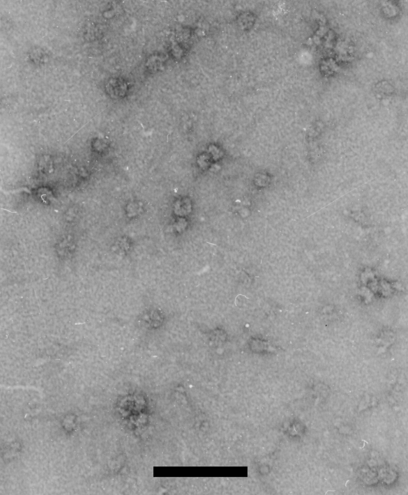
Salmonella cells were lysed by freezing and thawing. The ribosomes were separated from other soluble cellular components by centrifugation through a linear sucrose gradient. Gradient fractions containing ribosomes were dialyzed to remove excess sucrose. The field show here contains single 70S particles (lower left and right) as well as small polysomes consisting of 2, 3, or 4 70S particles attached by a single mRNA (top and middle right). The scale bar is 100 nm.
Electron microscopy was performed by Patricia Reilly at the Biomedical Electron Microscopy facility at MIT.