The Neural
Control of Visually Guided Eye Movements
B. Subcortical Mechanisms of Visually Guided Saccadic Eye Movements
 Neurons
in the upper layers of the superior colliculus have well-defined receptive
fields. Neurons in the deeper layers discharge prior to and during eye
movements; some of these cells also have visual receptive fields. Electrical
stimulation here elicits saccades at low currents. The basic operational
principle of this structure is shown in Figure
5. Schematized are three microelectrode recording sites; shown
are the receptive field locations of the neurons at the tip of each site.
Electrical stimulation at these locations results in a saccadic eye movement
that shift the center of gaze to the place where the receptive field had
been located prior to the eye movement. The basic process therefore involves
the computation of the retinal error signal between the present and desired
gaze direction; the signal so generated results in a saccade that nulls
the retinal error. The code utilized is called a retino-centric code. Neurons
in the upper layers of the superior colliculus have well-defined receptive
fields. Neurons in the deeper layers discharge prior to and during eye
movements; some of these cells also have visual receptive fields. Electrical
stimulation here elicits saccades at low currents. The basic operational
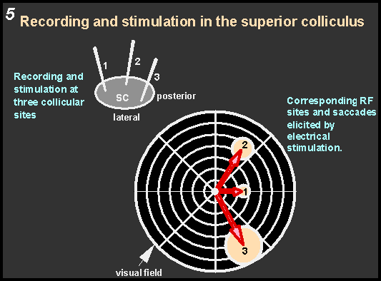
principle of this structure is shown in Figure
5. Schematized are three microelectrode recording sites; shown
are the receptive field locations of the neurons at the tip of each site.
Electrical stimulation at these locations results in a saccadic eye movement
that shift the center of gaze to the place where the receptive field had
been located prior to the eye movement. The basic process therefore involves
the computation of the retinal error signal between the present and desired
gaze direction; the signal so generated results in a saccade that nulls
the retinal error. The code utilized is called a retino-centric code.
|