
The human brain has roughly 10 billion neurons that communicate with one another through synaptic connections. Each neuron is capable of making 5,000-10,000 synapses its targets, leading to 50-100 trillion synaptic connections in the brain. The connections between neurons give rise to functional neural networks that provide the cellular substrate for higher cognitive functions such as learning, memory, and, ultimately, consciousness. How these complex connections are established during the early phases of nervous system development remains largely unknown.
Comparisons among immature and mature nervous systems have shown that individual neurons make a large number of supernumerary connections during early development. Correlated neural activity initiated by external stimulation then sculpts the pattern of connections through a selective strengthening of appropriate sets of latent connections, while eliminating the inappropriate. We have carried out experiments to study functional maturation of synaptic connections during early development.
Synaptic transmission
at newly formed glutamatergic synapses that contain postsynaptic NMDA receptors
is functionally "silent." Our laboratory has found that these "silent"
synapses contain functional AMPA receptors postsynaptically, but are incapable
of taking up endocytotic dyes presynaptically, suggesting that the lack of
synaptic transmission might derive from presynaptic deficiencies. Careful
analyses of the kinetics of AMPA and NMDA receptor activation revealed that
NMDA receptor-mediated currents could be activated selectively at synapses
containing both AMPA and NMDA receptors, under conditions where the flux of
neurotransmitter was reduced. We are exploring the pre- and postsynaptic mechanisms
that could influence the flux of the transmitter, including the presynaptic
vesicle fusion process during synaptic transmission. Interference with synaptobrevin,
a critical molecule for regulating vesicular fusion, converts a functional
into a "silent" synapse. These results suggest that one aspect of
the maturation of synaptic transmission is an enhancement in the flux of the
transmitter delivery in the synaptic cleft.

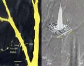
Activation of Synaptic Receptor in a single Synapse. Left, Visualization of functional presynaptic terminals by FM dye staining. Right, AMPA receptor map. White dots indicate the location at which glutamate was applied to the dendrite. The glutamate-evoked current obtained from each point is shown in the white traces above the white dots. The distance between the white and yellow dots represents the peak amplitude of the glutamate-evoked current at the corresponding white dots.

Associate Professor, Departments of Brain and Cognitive Sciences and Biology
Investigator, RIKEN-MIT Neuroscience Research Center
Guosong Liu
received his MD from Chuanbei Medical School in China, and his Ph.D. from
the University of California, Los Angeles. After postdoctoral training at
Stanford University, Dr. Liu joined the Department of Brain and Cognitive
Sciences, and the Picower Center for Learning and Memory at MIT in 1996. He
is a recipient of the Ursula Mandel Fellowship, the Outstanding Science Graduate
Student award from Sigma Xi and held the Poitras Career Development Professorship
at MIT.
laboratory webpage
 |