Molecular Walls Reveal Nano-Secrets of Cartilage
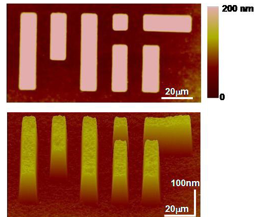
The image above were prepared by Department of Materials Science and Engineering Ph.D. student Lin Han (linhan@mit.edu) who is a joint student in the Ortiz and Grodzinsky research groups. The image was taken in an aqueous environment with a powerful microscope called an atomic force microscope (AFM) which uses a fine probe tip as nanosized "fingers" to "feel" surface contours and create a topographical imagMole (analogous to a record player). The sample shown was prepared by a method called "microcontact printing" which creates micrometer-scale molecular patterns. In this case, a layer of cartilage molecules called aggrecan were chemically end-attached within the MIT letter surface areas (height ~ 200 nm or one molecule thick!) and smaller molecules called a self-assembling monolayer (height ~ 1 nm, again one molecule thick!) are end-attached to the surface areas outside of the MIT letters. Aggrecan is thought to a primary contributor to the biomechanical properties of cartilage tissue, and its loss from the tissue is one of the early events in osteoarthritis and joint degradation. This pattern enables the direct visualization of the conformation and probing of the stiffness of these molecules at the nanoscale under near-physiological solution conditions. For more information : see Dean, et al. Macromolecules 2005. Download the PDF HERE. This research was funded by NIH Grant AR33236, NSF-NIRT 0403903, and the Whitaker Foundation.
Ortiz Nanomechanics Laboratory (Department of Materials Science and Engineering) |

This page was created and is updated by Christine Ortiz. Last Update : 04/24/05 Copyright 1999, Christine Ortiz, all rights reserved.