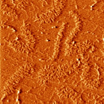
Tapping mode AFM image of a high density surface of aggrecan molecules in air
on mica.
Cartilage is a highly specialized, dense connective tissue found between the surfaces of movable articular joints whose main function is to bear stresses during joint motion. This complex biocomposite possesses high stiffness, toughness, strength, resiliency, and shock absorption. The extracellular matrix of cartilage is composed of many different molecules and structures. The negatively charged, disaccharide chondroitin sulfate glycosaminoglycan (CS-GAG) macromolecules are a major determinant of the tissue's ability to resist compressive and shear loading in vivo (e.g., responsible for >50% of the equilibrium compressive elastic modulus under normal physiological conditions (0.15 M salt concentration)). Approximately 100 CS-GAGs are covalently bound at extremely high densities (~2-4 nm separation distance), to a 250 kDa core protein forming the aggrecan molecule. The high charge density of the CS-GAGs of conjunction with their close packing cause these polysaccharide chains to take on a rod-like conformation rather than a random coil. Multiple aggrecan molecules self-assemble further to form supramolecular proteoglycan aggregates by non-covalently attaching to a hyaluronic acid or hyaluronan (HA) central filament, an interaction that is stabilized by the adjacent binding of a small glycoprotein called a link protein. These aggregates form the gel-like component of cartilage that is enmeshed within a network of reinforcing collagen fibrils. The structure of cartilage is
shown schematically here.
In this project, we are using the Atomic Force Microscope to image the conformation of the molecular constituents of cartilage aggregating proteoglycans from the length scale of the aggregate (~microns) down to the level of the individual glycosaminoglycan chains (~nm) in an ambient environment, as well as aqueous solutions of varying ionic strength, i.e. physiological and nonphysiological conditions.
Up until now, only 2-dimensional electron microscope images of prepared, fixed, and dried proteoglycan and aggrecan have been obtained. The atomic force microscope (AFM) offers a number of advantages including fluid imaging, lateral resolutions of less than one nanometer, minimal sample preparation and time (samples do not need to be coated, stained, or frozen), and the ability to use complementary techniques which provide information on other surface properties, such as stiffness, hardness, friction, or elasticity, in addition to topography. Using AFM, we were able to directly visualize the domain structure of individual aggrecan molecules and the variations in aggrecan conformation as a function of ionic strength, as well asa with age (fetal versus mature). As expected, a large increase in height was observed when the aggrecan monomers were fully hydrated at low ionic strength. As the ionic strength was increased, the counterions shielded the negative charges of the GAG chains, thereby reducing the Debye length and collapsing the molecule. The self-assembly process of aggrecan and hyaluronan to form aggregate was also directly observed. Ongoing experiments include studies of effects of ionic strength and pH on aggrecan conformation, characterization of the kinetics of the aggregate self-assembly process, and the use of specific G1- and G3- antibodies to further confirm aggrecan structures visualized by these methods. The nanomechanical properties of the individual
aggrecan molecules as a function of ionic strength and pH will be studied and
compared to the thesis project running in parallel on the
nanomechanics of glycosaminoglycan
biomimetic polymer brushes.