Nanoscale mapping of bio-molecular building blocks of brain
Complex biomolecular machineries enable our life processes and thus, understanding their nanoarchitecture can provide fundamental insights into biology. We showed that due to the crowded biological environment, these biomolecular nanostructures can remain invisible to existing technologies.
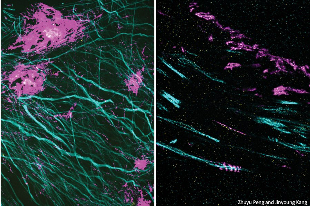
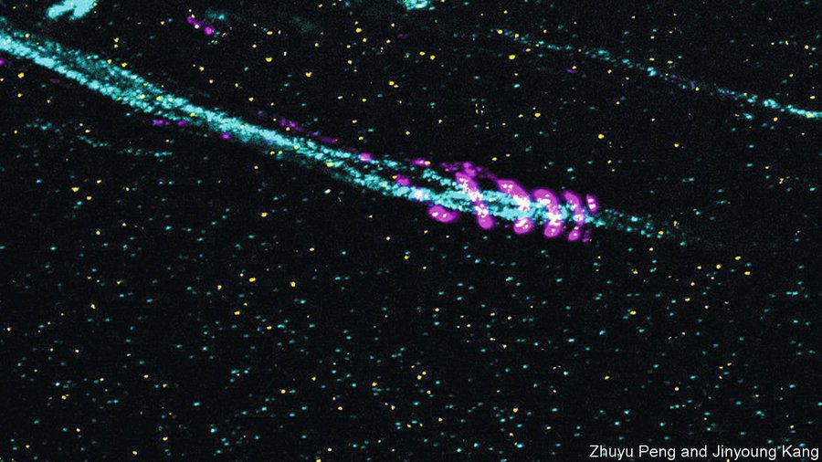
Hence, we developed a technology called expansion revealing (ExR) Microscopy, which decrowds the complex biomolecular environment and showed that it enables the discovery of fundamentally new nanostructures within biological specimens [D. Sarkar et. al., Nature Biomedical Engineering (2022)]. To achieve this, we developed an expansion mechanism of tissue-polymer hybrids that leverages both electrostatic and mechanical expansion to achieve high expansion factors with retention of the original biomolecules enabling decrowding effect. As examples of its capabilities, we illustrated in intact brain tissue the alignment of presynaptic calcium channels with postsynaptic machinery, which may facilitate precision synaptic transmission. We also revealed for the first time, the existence of periodic amyloid- beta nanoclusters colocalizing ion channel proteins in Alzheimer’s model mice, which may help generate novel hypotheses for Alzheimer’s pathology and neural excitability. Both these nanostructures were otherwise invisible without ExR even when imaged at the same level of super-resolution. Thus, the decrowding power of ExR is able to reveal novel nanostructures within intact brain circuitry and may find broad use in biology and medicine for unmasking nanostructures of importance in normal functions and disease.
We have also developed the technology to achieve highest expansion factor, reported till date (100- fold linear expansion), of tissue-polymer hybrids [D. Sarkar et. al., Society for Neuroscience (2016)]. Such high physical expansion factors allow imaging of biological specimens at sub-10 nm resolution (i.e., 300 nm (diffraction limit) / 100 (expansion factor)), using conventional diffraction limited microscopes.
We have shared this technology with dozens of labs worldwide, who are employing it to understand nanoscale architecture in brain and other biological structures as well as neurological disorders such as Alzheimer’s, Autism, Schizophrenia and Parkinson’s diseases.