| Summary |
Although mammalian galectins lack conventional signal sequences, they reach the cell surface by a novel mechanism and bind to glycoconjugates in on the plasma membrane and in the extracellular matrix (Barondes et al., 1994). The galectins consist of globular galectin-type CRDs with relatively minor accessory domains (
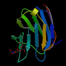
Cooper and Barondes, 1999) . A galectin-type CRD comprises a/b sandwich similar in overall topology to the L-type CRDs. However, the lack of sequence similarity and the different way in which sugar-binding sites are constructed in these two families of domains suggest that this topological similarity results from convergent evolution. Most galectins contain multiple sugar-binding sites, due to the presence of two galectin-type CRDs in a single polypeptide or as a result of dimerisation. A common function of the galectins may be to crosslink N-acetyllactosamine-containing structures found at cell surfaces and in the extracellular matrix.
Comparison of all the galectin sequences reveals conservation primarily of inwardly facing hydrophobic residues in b strands in the b-sandwich of the galectin fold (
Lobsanov et al., 1993;
Liao et al., 1994;
Leonidas et al., 1998). Eight residues that form the galactoside-binding site are conserved in most mammalian galectins, although a vertebrate galectin containing only six of the canonical galactose-binding residues interacts with mannose rather than galactose (
Swaminathan et al., 1999).
Galectin-3 (also known as MAC-2 antigen; CBP-35 or IgE-binding protein), a 35 Kd lectin which binds immunoglobulin E and which is composed of two domains: a N-terminal domain that consist of tandem repeats of a glycine/proline-rich sequence and a C-terminal galaptin domain. Galectin-3 has been shown to modulate apoptosis by interactions with bcl family members.
|