HST.563
LAB 4: Echocardiographic Imaging
Robert Manzke, Ph.D.

Contents
a. Technical principles
of ultrasound
b. Clinical aspects of
echocardiography
This lab is an introduction to
echocardiographic imaging. Echocardiography is a non-invasive (transthoracic
echo,TTE) or minimally-invasive (transesophageal echo,TEE; intracardiac echo,
ICE; intravascular ultrasound, IVUS) imaging technique which allows for
real-time visualization of the cardiac anatomy and function. Echocardiographic
imaging is the clinical standard for functional assessment of the heart and is
the firstline modality for assessing heart anatomy, structure, and function. It
provides a wealth of essential clinical information such as the size and shape
of the heart, its pumping capacity (Ejection Fraction, EF), as well as the
location and extent of any damage to its tissues. Due to the real-time
character and fine spatial resolution of ultrasound, it is especially useful
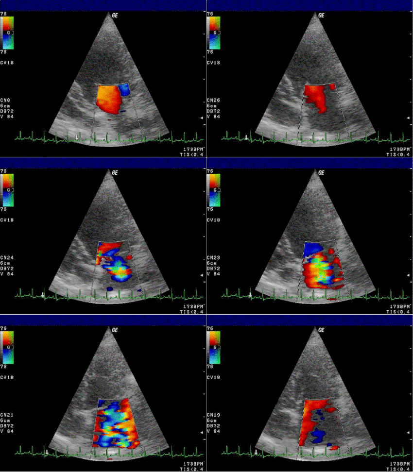
for assessing diseases of the heart valves. Doppler imaging techniques allow
physicians to detect abnormalities in the pattern of blood flow, such as the
retrograde flow of blood through leaky heart valves, known as regurgitation. By
imaging the motion of the heart wall, echocardiography can help detect the
presence and assess the severity of coronary artery disease and myocardial
infarcts. Echocardiography is also used to detect pathologies such as
hypertrophic cardiomyopathy in which the walls of the heart thicken in an
attempt to compensate for heart muscle weakness. A limitation in 2-dimensional
ultrasound is that the true three-dimensional anatomy is not captured.
Recently, new 3-dimensional ultrasound imaging techniques have been introduced,
which give a more complete picture of the anatomy in its entirety.
In preparation for this lab, you
will read three review papers on echocardiography [1-3] which summarize recent technological and clinical developments. Focus
on [1]. For further reading, we recommend
[4] for technical aspects of
ultrasound imaging and [5] for the clinical applications, as
well as material readily available on the web.
a. Technical principles of ultrasound
Ultrasound waves used in medical applications are primarily longitudinal waves
of mechanical pressure, which propagate in a medium such as human tissue
(though more recently, medical research into shear waves for ultrasound-based
strain imaging has also been extensive). The wave equation for pressure fields
in liquids and gases is given by
![]()
where pressure is given by ![]() , the density of the medium is represented by
, the density of the medium is represented by ![]() , the time by t and the bulk modulus by
, the time by t and the bulk modulus by ![]() .
.
The wave propagation velocity is  where
where ![]() is the wavelength and
is the wavelength and ![]() is the frequency of the pressure wave. The sound impedance is defined as
is the frequency of the pressure wave. The sound impedance is defined as ![]() ,
,
which represents the ratio of the stress force per unit area to the
displacement velocity (Table
1 shows typical values for different biological
tissues).
The intensity reflection and transmission factors are given by the expressions
 .
.
The ratio
of a reflected signal to the source signal is often expressed in decibels ![]()
Table 1: Ultrasound parameters.
|
Material |
Density in kgm-3 |
Propagation velocity in m/s |
Impedance in |
|
Water |
1000 |
1480 |
1.48 |
|
Muscle |
1080 |
1580 |
1.7 |
|
Fat |
900 |
1450 |
1.3 |
|
Brain |
1050 |
1540 |
1.6 |
|
Blood |
1030 |
1570 |
1.58 |
|
Bone |
1850 |
3500-4300 |
6.5-8.0 |
|
Air |
1.3 |
330 |
4´10-4 |
Note the very large differences between the soft tissues
and bone and air.
Signal
reflection factors at boundaries between soft
tissues are small:
e.g. fat
to muscle ![]() .
.
Reflection
factors between soft tissues and bone or
air are large:
e.g. bone
to brain ![]() .
.
The
intensity of the ultrasound wave decreases exponentially with the penetration
depth. The attenuation coefficient ![]() can be used to determine the total attenuation in dB/cm in
the medium according to
can be used to determine the total attenuation in dB/cm in
the medium according to
![]()
At an
ultrasound frequency of 1MHz, the attenuation is on the order of 1 dB/cm for
soft tissue such as kidney or liver (see Table
2), whereas for bone the coefficient is in the order of
20 dB/cm. The spatial resolution is dependent on the wavelength. A detectable
echo occurs if the structural distance is at the order of half the wavelength.
Table 2: Penetration depth and resolution in soft tissue.
|
Frequency
f in MHz |
Approximate
wavelength in mm |
Depth
in cm |
Typical
application |
|
3.5MHz |
0.44 |
15 |
Liver,
Heart |
|
7.5MHz |
0.21 |
7 |
Prostate |
|
15MHz |
0.1 |
1 |
Intravascular
applications |
In a
practical setup, ultrasound waves are produced and detected by piezoelectric
crystals, which change dimensions depending on an applied alternating
electrical field. In the simplest setup, a single transducer element transmits
a pulse into the tissue (see Figure
1). Immediately after transmission, the transducer is
switched to receive mode and reflected waves are detected, forming an image.
This simplistic setup is used for 1-dimensional A-mode imaging.

Figure 1: Simple A-line one element setup.
If a time
series of A-mode images is recorded, an M-mode image is obtained (see Figure 2).

Figure
2: M-mode image of the heart. Upper row shows
A-lines.
If the
single element transducer is physically swept along the one spatial axis, one
can form a 2-dimensional image (B-mode, see Figure
3).
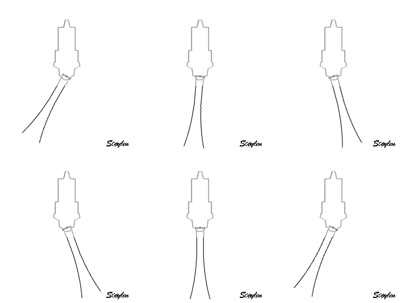
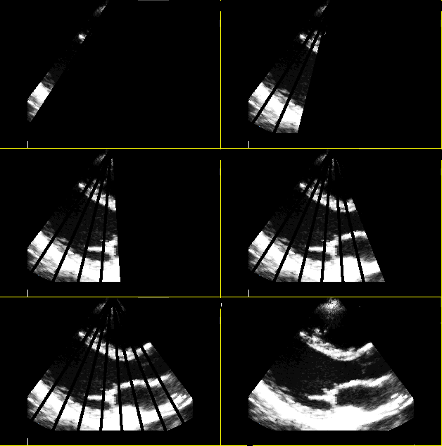
Figure
3: Mechanical movement of the single element
transducer (B-mode).
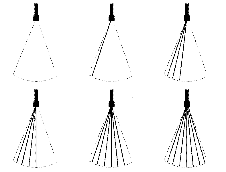
In most
cardiac applications today, electronically-steered phased arrays are used to
steer the focus of the ultrasound transducer to sample a 2D image slice through
the volume.
Such phased arrays typically consist of several piezo-electric elements
organized into a one-dimensional array. Each element of the phase array can be
pulsed with a certain transmission delay. This way, the focus of the resulting
wave field of all elements can be swept around a scan sector as shown in Figure 4.
Note that the smaller the imaging sector is, the faster echoes can be received
and, hence, the higher the frame rate and temporal resolution is. Likewise, the
shorter the imaging depth, the higher the pulse repetition frequency, the
higher the frame rate and associated temporal resolution.
If the phased
array is extended into a two-dimensional plane of elements, the focus can be
steered in three dimensions to sweep a conical volume. This is the basis for modern 3-dimensional
ultrasound transducers.

