(From http://webvision.med.utah.edu/VisualCortex.html)
The Primary Visual Cortex
by Matthew Schmolesky
The relationship between vision and the eye must have been correctly understood from the earliest times of human existence. Surpassing this most basic understanding, however, required careful dissection of the eye, optical fibers and brain structures of animals and/or humans. One view holds that the religious and social doctrines of the Pre-Classical civilizations (e.g. Egyptians, Sumerians, Assyrians, Babylonians, Hindus, Chinese, Indians, etc.) did not permit such dissections. The more primative herdsman and farmers surrounding the Mediterranean Sea did not have such inhibitions and are believed to have studied basic anatomy, albeit nonsystematically, during sacrificial rituals. These herdsman and farmers evolved a terminology for the anatomical structures they discovered.
Polyak (1957) maintains that this terminology was quickly adopted by the Greeks
of 600-400 B.C. when the study of anatomical organization was rigorously pursued.
During this time, physicians such as Hippocrates (460-380 B.C.), Alkmaeon (520
B.C.), and Anaxagoras (500 B.C.) carried out dissections of animal and human
bodies and brains and held opinions of some accuracy concerning brain function,
such as movement, sensation and thinking.
The height of Greek medical scientific knowledge was reached during the Hellenistic
or Alexandrian period from 323 to 212 B.C. The most prolific of medical scientists
was the "Prince of Physicians" Galen (129-201 A.D.) of Pergamon (present
day Turkey). As noted by Polyak (1957), Galen's contributions to the understanding
of human anatomy and physiology under normal and pathological conditions were
vast, and provided a foundation for medicine in the Arab period and, subsequently,
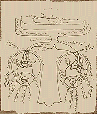
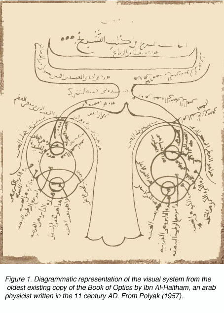
the Revival in Europe during the late Middle Ages and Modern Times. Figure 1
shows the arab adaptation of Galen's ideas on the pathways of the visual sytem
from eyes to forebrain.
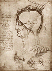
Though Galen accurately described much of the visual system gross anatomy, he did not recognize the decussation of fibers at the optic chiasm, nor did he trace the fibers to the dorsal lateral geniculate nucleus (LGNd) of the thalamus (Fig. 1). Instead, he suggested that they were connected to the lateral ventricles (which he named the "thalami"). This belief was consistent with the prevailing view on nerves in general at the time, namely that they were hollow channels carrying various "spirits" such as the visual spirit. Over one thousand years later, the beliefs of humanist scientists such as Leonardo Da Vinci (who pictorally described the same optic fiber to lateral ventricle pathway) (Fig. 2) and Des Cartes (who promoted the view of nerves carrying animal spirits) echoed Galen's teachings and demonstrate how little progress had been made in such a long time.
Following the Hellenistic period, the ethics supported during the first several
centuries of the Christian Era denied medical practitioners and scientists the
right to dissect human corpses. This stricture was expanded in the Dark Ages
to include the dissection of animals and, from this point until the 1600s, healers
were forced to rely almost entirely upon what little information they could
glean from the Classical period. Figure 3. Binocular stereoscopic visual system
as imagined by Des Cartes. The two retinal images of the arrow are accurately,
point for point, projected upon the surface of the cerebral ventricles and thence
to the centrally located pineal gland, H, the supposed 3seat of imagination
and common sense.