| Figure 1
Nature Cell Biology
1, 493 - 499 (1999)
Published online: 8 November 1999; | doi:10.1038/70281
Cooperative symmetry-breaking by actin polymerization in a model for
cell motilityAlexander van Oudenaarden
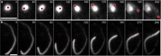
& Julie A. Theriot | | | | Figure 1. Symmetry-breaking in a cloud of actin filaments.
Time sequence of combined phase-contrast and fluorescence microscopy images
of an actin cloud that spontaneously breaks symmetry (a), and a bead
propelled by polymerizing filaments after symmetry-breaking (b). The
bead shown in b moves with a constant velocity of 0.12 ‘şm s
−1. The grey scale shows the fluorescence intensity of rhodamine-labelled
actin. The actual position of the bead is monitored using phase-contrast microscopy
and is represented by the red circle. The scale bars denote 5 ‘şm. Time elapsed
is shown in seconds.
|
| | | |  |
|