To view this video please enable JavaScript, and consider upgrading to a web browser that supports HTML5 video

To view this video please enable JavaScript, and consider upgrading to a web browser that supports HTML5 video

To view this video please enable JavaScript, and consider upgrading to a web browser that supports HTML5 video

Unraveling the Mysteries of the Brain
Our Mission
Our mission is to understand the brain and to apply that knowledge to improve human health and well-being. To accomplish these goals, we study the brain at many levels, across multiple disciplines, and we collaborate with academic, clinical, and industry partners around the world to challenge and probe the unknown.
More about us
From neurons to learning and memory
Mark Harnett investigates how electrical activity in mammalian cortical cells helps to produce neural computations that give rise to behavior.
To view this video please enable JavaScript, and consider upgrading to a web browser that supports HTML5 video

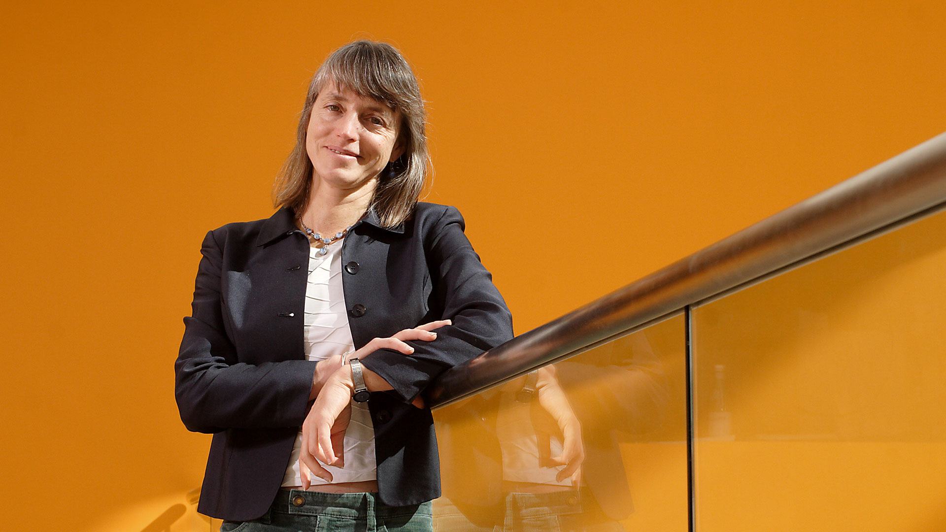
Nancy Kanwisher
Nancy Kanwisher studies the functional organization of the human brain as a window into the architecture of the mind.
Our Researchers
Our faculty consists of leading researchers with cutting-edge expertise and complementary approaches to understanding the brain.
See all Researchers