|
Singapore-MIT Alliance for Research & Technology |
||||||||||||||||||||||||||
BioSyM IRG
Publications Useful Links Global Enterprise for Micro Mechanics and Molecular Medicine (GEM4): www.gem4.org Mechanobiology Institute: http://mbi.nus.edu.sg/ Singapore-MIT Alliance for Research and Technology (SMART): http://smart.mit.edu/home.html
|
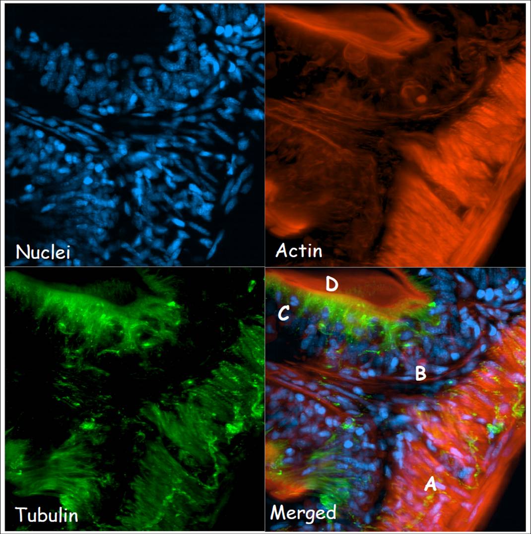
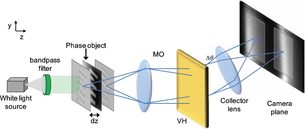
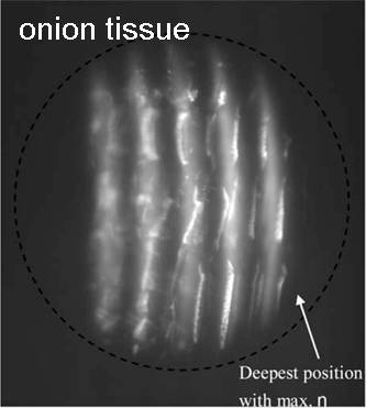
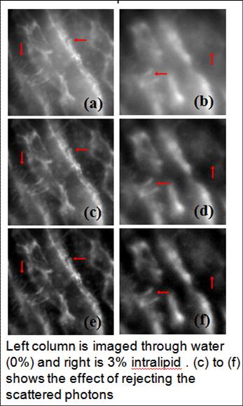
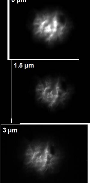
Research Projects Under Thrust 4: In-Vivo Cellular Systems EngineeringThrust 4 represents our explicit recognition of the need for our work to move toward in vivo applications. Current focus is on developing a range of imaging technologies. Several breakthrough imaging technologies are at various stages of development in BioSyM. Multi-plexed volume holography methods are under development that will apply rapid, intrinsically three-dimensional capabilities to biological imaging. In separate studies, a new index for liver fibrogenesis, Fibro-C, has been developed that enhances the diagnostic capabilities of standard histopathology. New methods are under study that would allow similar measures to be made in vivo, under minimally-invasive conditions.1. Novel optical microscopy and phase recovery methods for bioimaging(George Barbastathis (MIT/SMART-BioSyM), Peter T. C. So (MIT/SMART-BioSyM), Roger D. Kamm (MIT/SMART-BioSyM), H. Harry Asada (MIT/SMART-BioSyM), Paul Matsudaira (NUS), Colin J. R. Sheppard (NUS), Dipanjan Battacharya (SMART-BioSyM), Se Baek Oh (MIT), Justin Wu Lee (MIT) and Laura Waller (MIT))This project aims to develop non-invasive methods of imaging and phase recovery for imaging cells and tissue in vivo or in micro-biofluidic systems. We have developed optical instruments based on (1) scanning light-sheet microscopy, (2) volume holographic pupil microscopy, (3) colour transport of intensity. These provide three-dimensional image information in real time and with high resolution. Regeneration of intestinal epithelial tissue is a complex dynamic process that involves division and differentiation of stem cells at the base of the villi, migration towards the tip and apoptosis at villi tip. But the basic dynamics of this serial process has not been studied directly. To understand how cells in the intestinal epithelium migrate and the interactions between molecular motors and structural proteins, we are developing a laser sheet based microscope and other novel optical methods such as Multiplex-Volume holographic microscope to achieve improved spatio-temporal resolution. 1. Development of Laser-sheet based microscope for Bioimaging: We have built a SPIM (Selective Plane Illumination Microscopy) for live small animal imaging and are working on developing microscopic techniques towards live small animal imaging. In preliminary studies we are imaging with standard confocal approaches fixed intestinal tissue of mice, zebrafish and medaka fish model system to understand organization of different structural proteins forming intestine. Although confocal microscopy provides us a good x-y spatial resolution, but z-axis and temporal resolution are very poor. Also, confocal microscope has high photo-bleaching effect, not suitable for live small animal imaging. Using laser sheet based microscope we can excite a thin section of tissue (~micron) and collect the emission of this complete fluorescent plane simultaneously in a cooled CCD. We have built a laser sheet-based microscope and are working to further development of this technique for higher spatial resolution.
2. Development of Multiplex-Volume holographic microscope: We have started to build Multiplex volume hologram (MVH) microscope for spatial-spectral 4D imaging systems for real time imaging. In this method using a small multiplex volume holographic material in the collection pathway, we can spatially separate fluorescent emission from different fluorescent planes and simultaneously acquire spectral images of these planes in different parts of a cooled-CCD simultaneously. Using this method we can capture 3-D images of a specimen, simultaneously, without any scanning.
Selected publications
2. Transformation optics for cellular imaging and manipulation(Baile Zhang (SMART-BioSyM), George Barbastathis (MIT / SMART-BioSyM), Collin Sheppard (NUS), Hanhong Gao (MIT) and Satoshi Takahashi (MIT))Biological imaging at the molecular and cellular scales requires resolving of features in the order of 10's of nanometers. However, the fundamental diffraction limit discovered by Abbe in 1873 restricts the resolution of a conventional optical microscope to about half a wavelength (about 200 nm in air) due to the loss of high spatial frequency information carried in the evanescent waves. It is strongly desirable to develop a real time imaging technique that can provide subwavelength information. We propose to use gradient index (GRIN) dielectric materials to design and manufacture practical devices that can significantly improve the resolution of a common far-field microscope. Current simulations have shown that it is possible to achieve sub-100 nm resolution over broadband of visible spectrum by adopting this kind of device. Different from previous superlens and hyperlens that utilize surface plasmon resonance between metal and dielectric to capture subwavelength features within narrowband, our approach has great advantages (low-loss and broadband) and will be greatly simplified with existing micro-manufacturing technologies. Selected publications
3. Multi-scale imaging of liver fibrosis(Peter So (MIT / SMART-BioSyM), Hanry Yu (NUS/IBN), Jagath Rajapakse (NTU), Elijah Yew (SMART-BioSyM), Vijay Raj Singh (SMART-BioSyM), Daekeun Kim (MIT), Yan Jie (SMART-BioSyM), Jae Won Cha (MIT))Liver fibrosis is a wound-healing process in response to various toxic injuries and associated with almost all chronic liver
diseases.Inflammatory responses and excessive deposition
of extracellular matrix (ECM) are often observed with cirrhosis,
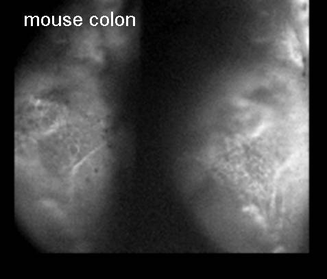
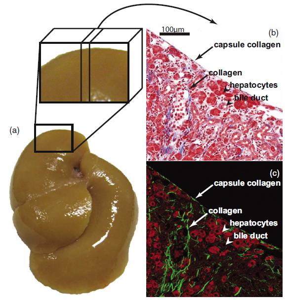
marking the end stage of fibrosis that is the major causes The goal of this project is to elucidate the molecular mechanism underlying fibrogenesis of the liver. This goal will be accomplished by cross-scale image quantification and informatics analysis. We study the complex pathological tissue transformation associated liver diseases such as fibrosis, cirrhosis, and cancer. High throughput optical imaging capabilities are being developed to quantify molecular and cellular features in disease models in 3D tissue models, mice and rats; and we correlate with data from clinical imaging modalities (MRI, ultrasound etc) to establish the biological basis of the diagnostic imaging of liver diseases (cancer, cirrhosis and fatty liver). We have shown that Second Harmonic Generation / Two-Photon-Excited Fluorescence (SHG/TPEF) imaging is a good substitute for Conventional Histological Imaging. A strong correlation between liver fibrosis progression on the anterior surface and the liver interior based on quantitative analysis of morphological features in both regions has been discovered. We are now one step closer to applying reflective or laparoscopic imaging of the liver surface to stage liver fibrosis, which can potentially be used clinically to complement or eventually replace the more invasive liver biopsy (J. Biomed. Optics, Vol. 15, 056007, 2010).
Comparison between histopathological staining and SHG/TPEF images. (a) A perfused fibrotic tissue was extracted and sectioned perpendicular to its surface to expose the liver boundary (inset in (a). (b) MT staining for a fibrotic tissue sample by the direct cutting is shown with collagen stained in blue, cytoplasm in red and cell nuclei in dark brown. (c) The SHG/TPEF image of the same sample is shown with collagen in pseudo green and cells in pseudo red. Features of the capsule collagen, collagen, hepatocytes, and bile duct shown in (b) the staining image can all be identified in (c) the SHG/TPEF image, respectively. (J. Biomed. Optics, Vol. 15, 056007, 2010) Selected publications
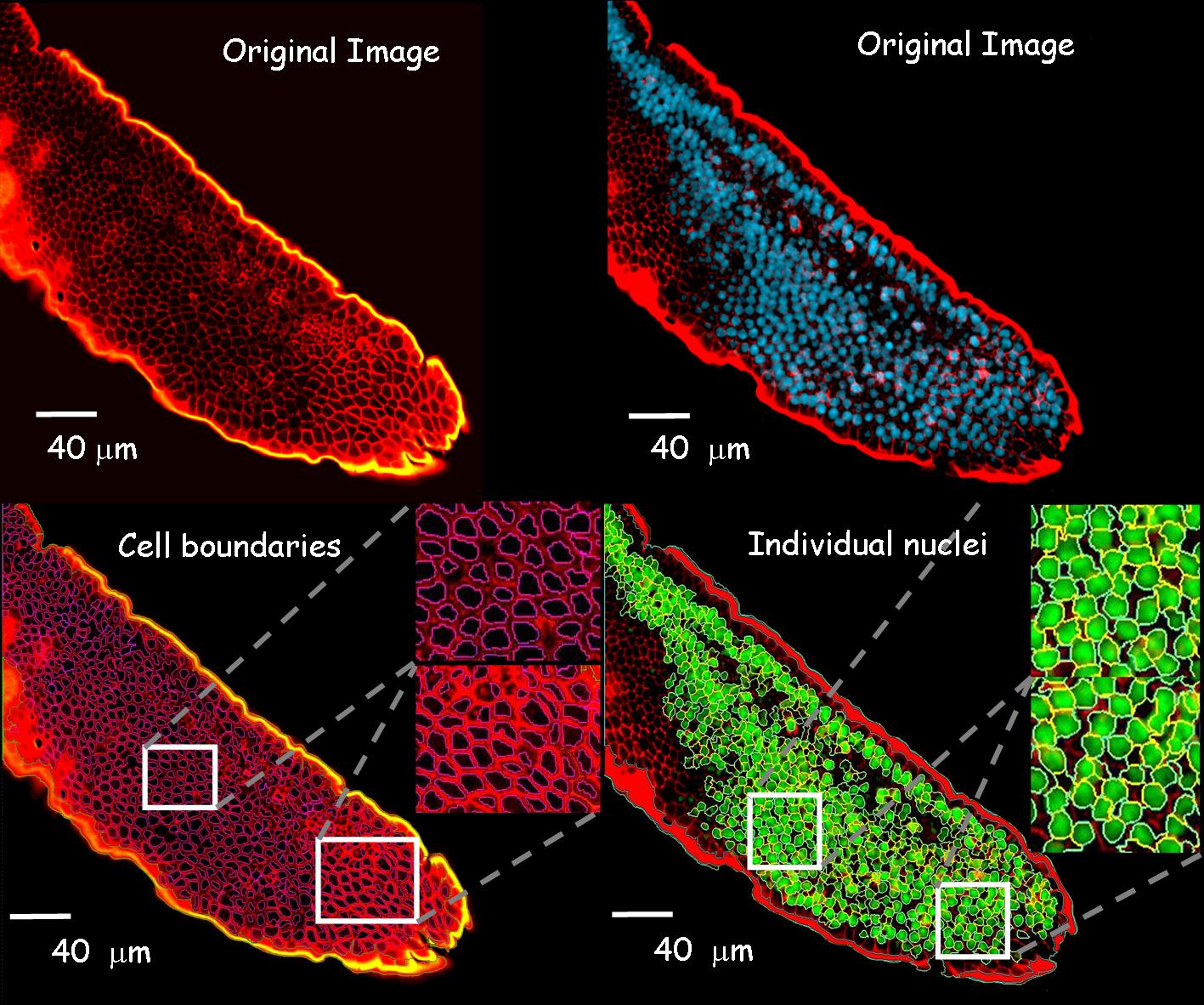
4. High Throughput-Image Cytometry Technologies(Peter So (MIT / SMART-BioSyM), Hanry Yu (NUS), Jagath Rajapakse (NTU), Colin Sheppard (NUS), George Barbastathis (MIT/SMART-BIoSyM), Roger Kamm (MIT/SMART-BioSyM), Elijah Yew (SMART-BioSyM), Vijay Raj Singh (SMART-BioSyM), Daekeun Kim (MIT), Yan Jie (SMART-BioSyM), Jae Won Cha (MIT), Heejin Choi (MIT))Commercial high numerical aperture microscope objectives are state-of-the-art optical instrument where monochromatic and chromatic aberrations are meticulously corrected. As many imaging and microfabrication applications require ever higher throughput, the imaging and manufacturing speed is limited by the object-side field of view (FOV) of these objectives that are typically less than 1 mm in diameter. The manufacturing of a large FOV microscope objective is difficult due to the fact that traditional spherical surfaces are insufficient to correct for monochromatic aberrations resulting in a need for aspherical surfaces. Large FOV microscope objectives require many large aspherical lenses that are difficult to fabricate and test leading to increased costs. At the same time the chromatic aberrations of the objective need to be carefully controlled using expensive glass types since the objective has to operate over a broad operating spectrum in both excitation and emission wavelengths. The goal of the project is to develop high throughput image cytometry technologies for cell in tissue matrix and in suspension. Here we are developing a novel adaptive optics based high-resolution and wide FOV objective for multiphoton microscopy with a FOV of of 7 mm x 7 mm. This system has the potential to greatly increase the throughput for many applications, including high-speed image cytometry for drug discovery as well as novel photolithographic nanofabrication for manufacturing microoptics, microfluidic chips, and tissue engineering scaffolds.
5. High Content Wide Field Excitation Techniques for Imaging, Spectroscopy, and Microfabrication(Peter So (MIT / SMART-BioSyM), Colin Sheppard (NUS), George Barbastathis (MIT/SMART-BIoSyM), Elijah Yew (SMART-BioSyM), Daekeun Kim (MIT), Partha Mondal (MIT))A collection of high content wide field imaging, spectroscopy and microfabrication techniques is developed for bio-applications. Super-resolution techniques in fluorescence mode was developed based on structured evanescence and plasmon waves with an extension for vibrational spectroscopy imaging. Temporal focusing nonlinear wide field excitation allows 3D-resolved image correlation spectroscopy, microrheology, and microfabrication.
|
|
||||||||||||||||||||||||