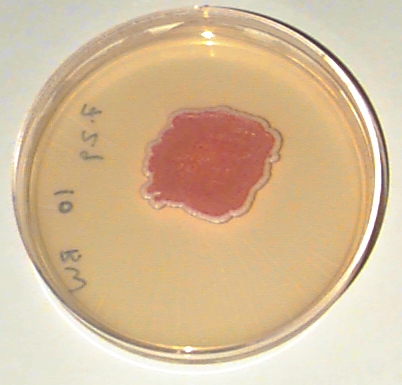
The photograph below shows a petri dish containing nutritive medium. In the center of the plate there is a patch of yeast cells. This patch has a red center and a white border. Based on this photo and the photos that follow, what can you conclude about why the patch has a red center and a white border? Please e-mail your answers, questions, and comments to: Brian White btwhite@mit.edu .
Figure 1: The original plate
Yeast cells from a specially-constructed yeast strain were suspended in water and 0.045ml of this suspension (roughly 100,000 cells, give-or-take an order of magnitude) was spread in a small puddle on the plate. When the water dried, the patch was a barely-visible cloudy area. The yeast suspension was well-mixed so that the cells were randomly dispersed in the patch.
The plate was incubated at 30C (86F) for 5 days. During this time, the yeasts went through many rounds of division, producung a dense patch of cells piled on top of and next to each other. As the cells get more and more crowded, the patch spread out a little (<5% in any direction). The result is shown below:
Yeast are single-celled microorganisms which grow
by dividing into two daughter cells every 1.5 hours or so (slower
if conditions are poor). The species used here, Saccharomyces
cerevisiae, is the same species as used in bread-baking. The
particular strain used here has been specially-constructed for
this experiment.
(1) Based on the above data, what models can you think of to explain the pattern of color?
Copyright 1996 Massachusetts Institute of Technology
Brian T. White
Thanks to:
Julie Archer - strains & lots of helpful advice.
Frank Solomon, Chris Kaiser, & Graham Walker - supplies
Sumati Murli - bench space & supplies