Harvard-MIT Division of Health Sciences and Technology
The Harvard-MIT Division of Health Sciences and Technology (HST) brings engineering, science, technology, and medicine to the solution of problems in biology and human health. A successful collaboration that spans more than 30 years between MIT, Harvard University, Harvard Medical School (HMS), and area teaching hospitals and research centers, HST is a pioneer in interdisciplinary educational and research programs designed to educate outstanding minds, cultivate leaders, create knowledge, and generate cost-effective preventive, diagnostic and therapeutic innovations. It is among the largest biomedical engineering and physician-scientist training programs in the United States.
Advances in biology and technology are bringing us to an era when diseases can be treated by "engineering" the phenotype of cells and tissues—when cell, tissue, and body functions can be manipulated using strategies that affect genes, cells, and their environment so they behave predictably. Advances in the diagnosis and prevention of disease are inexorably linked to these fundamental changes in our approach to disease management. Unquestionably, success in this area requires professionals with a broad range of skills that spans the domains of science, engineering, and medicine.
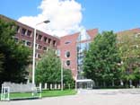 The Whitaker College of Health Sciences and Technology, HST's campus at MIT (Building E25). Photo by Mario Casal. |
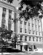 The Toteson Medical Center in Boston's Longwood medical area. Photo courtesy of Harvard Medical School. |
HST is dedicated to integrating these disciplines into an educational program that carries engineering and the physical and biological sciences from the laboratory bench to the patient's bedside, and, conversely, brings clinical insights from the bedside to the bench. HST's programs are committed to exploring the fundamental principles underlying disease, to seeking new pharmaceuticals and devices that ameliorate human suffering, and to training the next generation of physicians, scientists, and engineers to do the same. Thus, HST trains physicians with a deep understanding of the underlying quantitative and molecular science of medicine and biomedical research. HST PhD students similarly acquire a deep understanding of engineering and the physical and biological sciences. This unique training is complemented with hands-on experience in the clinic or in industry.
HST's administrative home is located at the Whitaker College of Health Sciences and Technology at MIT. As one of the five medical societies at Harvard Medical School, HST also maintains an office at the medical school's quadrangle campus in Boston. HST's two codirectors, Martha L. Gray for MIT and Joseph Bonventre for HMS, report to the provost and the vice president for research at MIT, as well as to the HMS executive dean for academic programs and the dean of Harvard Medical School. Richard N. Mitchell, assistant professor of Pathology at Harvard Medical School, serves as the division's associate director and director of student affairs.
Degree Programs
HST currently enrolls approximately 400 students who work with more than 200 faculty and affiliated faculty members from the Harvard and MIT communities. Eight multidisciplinary graduate degree options are offered, each targeted at students with different backgrounds and goals, each requiring a focused educational and research program, and each offering a different level of clinical training:
- Medical Sciences Program (MD)
- Medical Engineering and Medical Physics Program (MEMP)
- Speech and Hearing Bioscience Technology Doctoral Program (SHBT)
- Radiological Sciences Joint Program (RSJP)
- Medical Informatics Program (MI)
- Clinical Investigator Training Program (CITP)
- Master's of Engineering in Biomedical Engineering (MEMB)
- Biomedical Enterprise Program (BEP)
Research Programs
HST's research programs reflect a mix of cultures in applying the tools of medicine, engineering, and science to problems in human health and medicine. Research initiatives are conducted in three targeted focus areas:
- Biomedical Imaging
- Informatics and Computational Biomedical Sciences
- Regenerative and Functional Biomedical Technologies
and two crosscutting research programs:
- Speech and Hearing Bioscience and Technology
- Cardiovascular Sciences and Technology
Highlights
Events
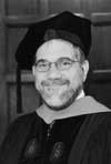 Steven E. Hyman, MD, delivered the keynote address at HST's 2003 commencement. Photo by L. Barry Hetherington. |
Steven E. Hyman, provost of Harvard University and professor of neurobiology at Harvard Medical School, delivered the keynote address at HST's graduation on June 6, 2003 at which 54 graduates received degrees (8 PhD degrees, 29 MD degrees, and 17 master's degrees). Ten MD students graduated cum laude. HST's graduating class of 2003 represented 14 states and 9 foreign countries.
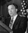 Mark B. McClellan, MD, PhD, (HST'89), and keynote speaker at the 16th annual Forum. Photo by L. Barry Hetherington. |
The 2003 HST Forum "FDA's Strategies for Improving Health Care" surpassed the number of research posters submitted for previous years (77) and featured keynote speaker Mark B. McClellan (HST'89), commissioner of the Food and Drug Administration. The 16th annual HST Forum welcomed more than 300 attendees on March 20 at the Harvard Club of Boston.
HST's annual John F. and Virginia B. Taplin Awards Symposium was held on May 22 and the philanthropists, for whom the awards are named, were in attendance. Presentations were given by the 2002 Taplin Fellows: Bertrand Delgutte, Richard N. Mitchell, Shamil R. Sunyaev, and T. Bernard Kinane.
In the fall of 2002, HST relaunched its web site, which enabled users to update personal information, RSVP for events, and submit events and news items. Additionally, authorized contributors can update the site's content directly though their web browser thereby improving the timeliness and accuracy of the content.
Dava Newman, associate professor of aeronautics and astronautics at MIT and HST affiliated faculty member, was first mate and education director of the Galeta Odyseey: World Contract project, an educational global outreach program.
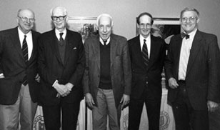 Walter H. Abelmann, MD, William S. Beck, MD, George B. Benedek, PhD, W. Hallowell Churchill, MD, and Dwight R. Robinson, MD, were presented with a special award to mark more than 30 years of service to HST. Photo by L. Barry Hetherington. |
George B. Benedict, Walter H. Abelmann, William S. Beck, Dwight R. Robinson, and W. Hallowell Churchill, were presented with special award to mark their more than 30 years of service to HST.
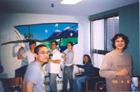 Austin Huang (MEMP), Xiaomin Mou (SHBT, and mural designer), Eduardo Torres-Jara (friend), Teresa Santos (SHBT), Jenny Mu (MEMP), and Felipe Jain (MD) Photo by Luwam Semere (MD). |
The third annual HST Community Service Day was held on Sunday, April 6 at the Barbara McInnis House, site of one of Boston's Healthcare for the Homeless Programs, in Roxbury. About 60 volunteers, including students, members of the faculty and staff, and friends participated.
Academics
HST was awarded a Neuroimaging Training grant by the National Institute of Biomedical Imaging and Bioengineering (NIBIB) to develop a curriculum that focuses on the application of modern brain imaging tools toward solving basic and clinical neuroscience questions.
A new training program in Neuroimaging, within the MEMP PhD program, was established with a training grant funded by the National Institutes of Health (NIH). The program was developed in direct response to an unmet need for greater numbers of skilled research scientists and academic leaders trained at the interface of imaging technology and clinical neuroscience. Dr. Bruce Rosen, the principle investigator of the grant, is the director of Massachusetts General Hospital's Nuclear Magnetic Resonance Center and the MGH/MIT/HST Athinoula Martinos Center for Biomedical Imaging. Dr. Rosen holds a joint HST/Harvard Medical School faculty appointment; he directs HST's Magnetic Resonance course, advises and mentors HST students, and is an active member of several core HST committees. The first trainees will be appointed to the training program in fall 2004.
The MD Admissions Committee received 546 applications. More than one-quarter (156) of the applicants were interviewed, and 43 of those were offered admissions. The PHD Admissions Committee received 243 applicants, and 30 will matriculate to HST's three PhD programs in September; and 21 students were admitted from a field of 50 to three HST master's degree programs. A total of 413 matriculated students were enrolled in HST programs in 2002–2003.
In collaboration with the Whitehead Institute/MIT Center for Genome Research, HST has launched a new diversity initiative aimed at increasing the number of underrepresented minority groups in the field of genomics.
New classes were added in 2002–2003, largely as a result of new programs added last academic year.
| HST.035 | Principles and Practice of Human Pathophysiology |
| HST.512* | Genomic Medicine |
| HST.527 | Blood Vessels and Endothelial Phenotypes in Health and Disease |
| HST.558J | Introduction to Modeling & Simulations |
| HST.569 | Biomedical Optics |
| HST.592 | Seminar in Computational Biology |
| HST.906 | Role of Physicians and Scientists in the Business World |
| HST.958 | Biomedical Information Technology |
| HST.971 | Building a Biomedical Enterprise: Strategic and Organizational Analysis |
| HST.977* | Critical Reading and Technological Assessment of Biomedical Devices |
| HST.975* | Clinical Trials in Biomedical Enterprise |
| HST.979* | Analysts' Club |
*taught under a special topics number pending approval of the permanent number.
Additionally, Evaluating a Biomedical Business Concept was offered as a special topic in Biomedical Enterprises.
Clinical training for MEMP students was expanded to include a second option at Massachusetts General Hospital (MGH). This option is targeted at students in the cellular and molecular track and is being offered by faculty with a clinical specialty in pediatrics.
HST established a student compliance system for four new federal regulations applicable to the clinical training of its students, including Health Insurance Portability and Accountable Act (HIPAA—a patient privacy law), Universal Health Precautions training on Blood Borne Pathogens, Occupational Safety and Health Administration (OSHA) regulated a mandatory fitting of respirator masks to protect against diseases such as TB and SARS, and mandatory TB testing.
HST formed ties with Cambridge University, UK, helping them to develop a program modeled after HST's Biomedical Enterprise Program (BEP). The focus of the HST-Cambridge curriculum development and collaboration has been a series of program-specific integrative subjects that focus on issues that fall within the interface of business and bioscience. Cambridge-MIT workshops and joint student projects have provided additional opportunities for students to interact with one another and with leaders in industry from both sides of the Atlantic.
HST collaborated with MGH, with funding from NIH and NSF, to provide a summer institute in Biomedical Optics. The program runs from June 11 to August 15, 2003, and a total of 12 undergraduate and graduate students are enrolled.
The MD Curriculum Committee approved a number of new initiatives this year. In response to a need expressed by HST MD students for additional mentorship, two new programs will be piloted in the coming year and will involve physicians from across the Longwood Medical Area. One program is intended for all HST MD students, while a second is specifically for fourth-year students facing internship decisions.
The Graduate Committee established a structure for reviewing several aspects of the PhD programs, and convened four subcommittees. The first committee is developing a unified theory for HST's various PhD programs, including issues related to qualifying exams and training grants. A second is reviewing the first year PhD program and making recommendations to improve the first-year experience. A third created a new generic HST course evaluation form, which was implemented in all PhD and some MD courses in spring 2003. The fourth delineated and approved a curriculum for the cellular and molecular track of the MEMP (Medical Engineering and Medical Physics) PhD program.
The HST MD Curriculum Committee reviewed the results of a fourth-year student survey developed in summer 2002 to gauge graduating students' view of the curriculum. One initiative that came out of the survey results was a mandate to incorporate communication skills training into the HST curriculum. Susanne Klingenstein, HST lecturer, will be creating and course and a workshop to further the HST communication curriculum.
HST began collaborating with MIT's OpenCourseWare project and anticipates having course materials for a number of HST courses online by September 30, 2003.
Faculty
New faculty appointments:
New joint faculty member Shamil Sunyaev arrived at the beginning of the academic year from the European Molecular Biology Laboratory (EMBL) in Heidelberg, Germany. Dr. Sunyaev's appointment as assistant professor of medicine at Harvard Medical School is pending review by the HMS Governing Board. He is based in the Department of Genetics at Brigham and Women's Hospital where he is focusing on the statistical analysis of genomic data from evolutionary and structural perspectives.
Affiliated faculty members Lucila Ohno-Machado, associate professor of radiology at HMS/BWH, and Gregory Sorensen, associate professor of radiology at HMS/MGH were appointed to the joint faculty.
New affiliated faculty members who joined HST in academic year 2002–2003:
- Ron Alkalay, instructor in orthopedic surgery, HMS, BIDMC
- Jerry Avorn, associate professor of medicine, HMS, BWH
- Brett Bouma, associate professor of dermatology, HMS, MGH
- Aziz Boxwala, assistant professor of radiology, HMS, BWH
- David Caplan, professor of neurology, HMS, MGH
- Johannes de Boer, assistant professor of dermatology, HMS, MGH
- Christopher Halpin, assistant professor of otology and laryngology, HMS, MEEI
- Stefan Heller, assistant professor of otology and laryngology, HMS, MEEI
- Robert Hillman, associate professor of otology and laryngology, HMS, MEEI
- James Bradley Kobler, assistant professor of otology and laryngology, HMS, MEEI
- Sharon Kujawa, associate professor of otology and laryngology, HMS, MEEI
- Michael J. McKenna, associate professor of otology and laryngology, HMS, MEEI
- Cynthia Casson Morton, William Lambert Richardson professor of obstetrics, gynecology and reproductive biology, HMS, BWH
- Omolola Ijeoma Ogunyemi, instructor in radiology, HMS, BWH
- Andrew Oxenham, principal research scientist, Research Laboratory of Electronics, MIT
- Brian Seed, professor of genetics, HMS, MGH
- Anne B. Skvorak-Giersch, instructor in pathology, HMS, BWH
- Guillermo James Tearney, assistant professor of pathology, HMS, MGH
- Staal Armund Vinterbo, instructor in radiology, HMS, BWH
- Qing T. Zeng, instructor in radiology, HMS, BWH
Faculty promotions:
- HST faculty member Bertrand A.R. Delgutte, PhD promoted to Senior Research Scientist in the Research Laboratory of Electronics' Auditory Physiology group.
- Joint faculty member Hugh Herr was promoted to assistant professor of physical medicine and rehabilitation at HMS/Spaulding Rehabilitation Hospital.
- Joint faculty member Lee Schwamm was promoted to associate professor of neurology at HMS. MGH. Dr. Schwamm was also named assistant director of HST's Clinical Research Center at MIT.
- Joint faculty member Mehmet Toner (HST PhD '89) was promoted to professor of surgery at HMS, MGH.
- HST affiliated faculty Nancy Andrews was promoted to professor of pediatrics at HMS and Children's Hospital.
- Affiliated faculty Barbara Fullerton was promoted to assistant professor of otology and laryngology, HMS, MEEI.
- Affiliated faculty Alan Moses, was promoted to professor of medicine at HMS/BIDMC/Joplin Diabetes Center.
Milestones
Student affairs coordinator Catherine Modica received the Laya W. Wiesner Award honoring Mrs. Wiesner's contributions to women's activities at the Institute. The award, established in 1980 by the MIT Women's League, is presented to the individual who has most enhanced MIT community life.
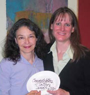 MIT institutional award winners and Cathy Modica and Patty Cunningham. Photo by Alison N. Haughton. |
Project manager Patty Cunningham won an 2003 Infinite Mile Award for her outstanding achievement, and was recognized for making extraordinary contributions in helping HST carry out its mission.
HST's Advisory Council convened on October 21 and March 20. The first meeting focused on biomedical imaging and neuroimaging research, and the second looked at creating a long-term strategic vision for HST's research endeavors.
HST faculty, staff, students, and alumni mourned the passing of:
- William S. Beck, MD, 79, professor of medicine, emeritus at Harvard Medical School and professor of health sciences and technology, emeritus at HST, passed away on Tuesday, May 27, 2003. Dr. Beck was the first director of HST.080 Hematology; and continued to lecture in the course until last year.
- Donald S. O'Hara, PhD, 67, passed away on February 2, 2003. Dr. O'Hara was a lecturer on biological chemistry and molecular pharmacology in the Department of Medicine at HMS. He was the codirector of HST.146 Human Biochemistry and Metabolic Diseases.
- William C. Quist, MD, PhD, 46, passed away on July 08, 2003. Dr. Quist was the associate director of the HST Human Pathology course for many years. In 1999, he was the recipient of the Irving London Teaching Award, which recognizes faculty who have made outstanding contributions to the training of HST students.
Alumni/ae
Alan D. D'Andrea (MD'83) is the senior author of a report of the discovery and cloning of six new genes responsible for inherited breast cancer. (NG Howlett et al., Science 2002; 606-609.)
Denis W. Choi (MD'78), executive vice president of neurosciences at Merck Research Laboratories in Whitehouse Station, N.J., spent an evening with HST students answering questions about the industry side of clinical research.
Steven P. Keller (PhD'02) was one of 11 Harvard graduate students and fellows who developed and taught basic science courses at a new medical school in Katmandu University.
Student Honors and Awards
Emanuela Binello, PhD, (MEMP), received MIT's Graduate Student Council Teaching Award for her work as teaching assistant for HST.201/202Introduction to Clinical Medicine and Medical Engineering.
Scott Boyd (MD/PhD) won the 2002 NF Research Prize given by the National Neurofibromatosis Foundation; his submission was on "Mammalian Synthetic Lethal Screens for NF1 Therapeutic Targets."
Michael R. Folkert, as a member of the Student Emergency Medical Services (SEMS) team, received MIT's William L. Stewart Jr. Award, which recognizes outstanding contributions by an individual student or student organization to extracurricular activities and events during the preceding year. Folkert was one of the founders of SEMS. This was Folkert's second Stewart Award.
Hertz Foundation Fellowships (continuing support): Lily Y. Kim, Andrew Levin, and E. Courtenay Wilson.
Howard Hughes Medical Institute award winners include: Robert Den, Sang D. Kim, and Mohammad Siddiqui. HHMI continuing support: David Berry, Christina Boulton, Kevin S. King, Stephanie Misono, Yvonne Ou, Ryuji Suzuki, David Ting, Vladimir Vinarsky, and Nikhil Wagle.
Courtney Lane (SHBT), won the first annual Helen Carr Peake Research Prize for her work on spatial release from masting. The Peake Research Prize is given to an MIT student for bioengineering research performed in either the Research Laboratory of Electronics or the Eaton-Peabody Laboratory of Research. Speech and Hearing Bioscience and Technology (SHBT) student Joshua Bernstein received an Honorable Mention for the Peake Research Prize for his research on the role of harmonic content in pitch perception.
Craig Lewiston (SHBT), received the 2003 HST Student Leadership Award, bestowed annually upon that student who contributes the most to the personal growth and professional development of his or her fellow students in HST.
National Science Foundation Fellowships (continuing support): Jose O. Aleman, Lauren J. O'Donnell, Laura C. Redi, Adam Rosenthal, Jocelyn E. Songer, and Lauryn R. Zipse.
David O'Gorman, (SHBT), was the recipient of a NIH Individual National Research Service Award.
Michael Pacold, first-year MD student, was awarded a Paul and Daisy Soros Fellowship for New Americans.
Kush Parmar (MD/PhD) won a Dean's Community Service Award from HMS for his work with the Cruz Blanca Initiative, a humanitarian effort in Mexico.
Whitaker Foundation Fellowships (continuing support): Gil Alterovitz, Aaron B. Baker, Erika L. Brown,
Steven K. Charles, David A. Eavarone, Paul Matthew George, Kevin R. King, Tony H. Ko, Sylaja Murikipudi, David N. Nguyen, Eric A. Osborn, Andrew G. Richardson, Christina E. Silcox, Joshua Tam, Juwell W. Wu, Peter I. Wu, and Ernest N. Yeh.
Faculty Honors And Awards
Richard J. Cohen, Whitaker professor in biomedical engineering, MIT was elected to the Association of American Physicians.
Jeffrey B. Cooper, PhD, affiliated faculty and associate professor of anesthesia, shared in the Janssen Elder Care Award.
George Q. Daley, assistant professor of medicine, HMS, MGH was elected to the American Society for Clinical Investigation.
Alan D. D'Andrea affiliated faculty and professor of pediatrics, HMS, CHMC was elected to the Association of American Physicians.
 Trudy M. Van Houten, PhD, (one of the recipients of the 2003 Irving M. London Teaching Award) with presenter Joaquin Blaya (MEMP). Photo by L. Barry Hetherington. |
Lee Gehrke, PhD, Herman von Helmholtz professor of health sciences and technology, MIT, and Trudy M. Van Houten, PhD, lecturer in anatomy, Program in Medical Education, and clinical instructor in radiology, BWH received the 2003Irving M. London Teaching Award. The London Award recognizes faculty who have made outstanding contributions to the training of HST students.
Robert S. Langer, Jr., ScD, Kenneth J. Germeshausen professor of chemical and biomedical engineering, MIT was elected to the Academy of Achievement and was the recipient of the 2003 John Fritz Medal, referred to as the highest award in the engineering profession, is presented each year for scientific or industrial achievement in any field of pure or applied science.
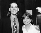
 Valerie J. Pronto-Stelluto, MD, (recipient of the 2003 Thomas A. McMahon Mentoring Award) with presenter Megan Hepler (RSJP). Photo by L. Barry Hetherington. |
Leonid Mirny, the Samuel A. Goldblith career development assistant of health sciences and technology and physics was a recipient of a 2003 Sloan Fellow. He was chosen from more than 500 nominees, young scientists nominated "on the basis of their exceptional promise to contribute to the advancement of knowledge" As 2003 Fellow, he will receive a grant of $40,000 for two years.
John Rosowski, associate professor of otology and laryngology, HMS was elected to the American Otologic Society.
Valerie J. Pronio-Stelluto, HST affiliated faculty and instructor in medicine at HMS and Mt. Auburn Hospital received the Thomas A. McMahon Mentoring Award, which is presented to a faculty member who not only provides scientific guidance to his or her students, but also inspires personal growth.
Educational Initiatives
VaNTH Engineering Research Center for Bioengineering Educational Technologies
A consortium of Vanderbilt University, Northwestern University, University of Texas at Austin, and HST, VaNTH is one of 20 engineering research centers (ERC) funded by the National Science Foundation. The ERCs are charged with "embodying NSF's strategic interests in the integration of research and education, in partnerships with industry, in the development of shared infrastructure, and in the improvement of science and engineering graduates' ability to contribute to National interests." Unique among the ERCs, the VaNTH initiative focuses on the educational enterprise. Its mission: to "unite educators and engineers, in industry and academia, to develop curricula and technologies that will educate future generations of bioengineers." Faculty and researchers in the VaNTH ERC are developing challenged-based learning modules that teach bioengineering content and provide an opportunity for the learner to explore the inter-connections and relationships among the selected concepts. VaNTH stresses inclusion of learning sciences, educational technology, and assessment, in both course module development and its related outreach programs. In the past year, VaNTH researchers in HST have developed, deployed, and evaluated instructional modules in the areas of spectral analysis, capillary filtration, renal physiology, respiratory biomechanics, and statistical analysis of fMRI data. Additional modules on the topics of microscale diffusion and endothelial biology are under development and will be used for teaching in the coming academic year. Other VaNTH efforts address the integration of biomedical ethics and communications into the curriculum.
BioMatrix
The goal of BioMatrix is to contribute to the professional and personal growth of MIT students by providing an opportunity for them to connect with mentors who can help them explore career decisions. Currently, BioMatrix has a membership of more than 170 MIT undergraduates, over 70 MIT graduate and Harvard medical students, and 70 mentors who are faculty, clinicians, researchers, and professionals in industry.
The structure of BioMatrix has three main components: monthly dinners organized by a student-led subcommittee around selected themes; student- or mentor-initiated small-group activities called BIOs ("BioMatrix Interactions on the Outside"), which include individual and small-group talks, visits, and professional and social opportunities; and a website with profiles of members and an event calendar. Mentors and students work together on programming, membership, communications, and assessment committees, which has built relationships across the membership categories and given student members opportunities to enhance their leadership skills. And as it moves into its fourth year, one of the goals of the program, for students to establish close relationships with faculty and other practitioner-mentors, is clearly being met: the first 'generation' of BioMatrix undergraduates is receiving useful advice as they approach graduate education or professional life. BioMatrix is becoming known in the MIT community—and in other higher education institutions—for its work in meeting the personal and professional needs of MIT students.
Biomedical Optics Summer Institute
In conjunction with the Wellman Laboratories of Photomedicine at Massachusetts General Hospital, HST offers a ten-week summer internship program to introduce undergraduate and graduate student participants to the field of biomedical optics — the use of light in biology and medicine. Supported by the NSF and the NIH, the program utilizes a combination of classroom study and laboratory research to offer participants preliminary exposure to a novel scientific field. In addition, communication and ethics training components and the formation of peer and mentoring networks broaden the scope of the program and enhance the students' summer research experience.
Bioinformatics and Integrative Genomics
HST in collaboration with the Whitehead Center for Genome Research is in the process of implementing a recruitment strategy for bioinformatics and integrative genomics (BIG), which targets and recruits underrepresented minorities. As a part of its recruitment strategy, HST will identify and develop relationships with faculty and administrators with relevant academic disciplines at universities and colleges to engage minority students. The HST diversity initiative is done through the domains of biomedical optics, genomics, bioinformatics, and bioengineering. The program will consist of a summer research experience, mentoring by faculty and peers, training in the ethical conduct of research, and technical writing and communication training.
Research Achievements
The research of the HST core faculty and research staff covers a wide spectrum of biomedical areas. In addition to laboratories at HST, MIT, Harvard University, and the Harvard Medical School, research collaborations include several HMS teaching hospitals in the Boston area (including Massachusetts General Hospital, Brigham and Women's Hospital, Beth Israel Deaconess Medical Center, Dana Farber Cancer Institute, and Children's Hospital).
Regenerative and Functional Biomedical Technologies
The objective of HST's efforts in regenerative biomedical technologies is the cost-effective replacement of cell, tissue, and organ function. Toward this end, HST researchers apply the rigors of physical sciences to problems in medicine and biology. In particular, this work seeks to understand, harness, and engineer tissues, cells, and molecules. A principle site of this effort is the Harvard-MIT Biomedical Engineering Center, located on the MIT campus. There, seven resident faculty members supervise a scientific staff of 90. An additional 70 affiliated faculty at MIT and HMS use the center's extensive facilities and resources, which support work in a broad range of integrated sciences from molecular and cell biology to animal physiology.
Elazer R. Edelman (HST'83) is the Thomas D. and Virginia W. Cabot associate professor of HST and director of the Harvard-MIT Biomedical Engineering Center. Dr. Edelman and colleagues in his laboratory use elements of continuum mechanics, digital signal processing, molecular biology, and polymeric controlled release technology to examine the cellular and molecular mechanisms that transform stable coronary-artery disease to unstable coronary syndromes. Tissue-generated cells, for example, deliver growth factors and growth inhibitors for the study and potential treatment of accelerated arterial disease following angioplasty and bypass surgery. The laboratory holds patents for drug-delivery devices, tissue-engineered implants, and new drug formulations.
Michael S. Feld heads the MIT Laser Biomedical Research Center, an NIH Biomedical Technology Resource housed in the Spectroscopy Laboratory, which develops basic scientific understanding, new techniques, and technology for advanced biomedical applications of lasers. Fluorescence, reflectance, near-infrared Raman scattering, light-scattering spectroscopy, and low-coherence interferometry are being used for histological and biochemical analysis of tissues, diagnosis and imaging of disease, and cell biology applications. Clinical studies are being conducted with researchers from the Mayo Clinic Foundation, the Brigham and Women's Hospital, Metrowest Medical Center, Beth Israel Hospital, and Boston University Medical Center. Clinical studies using trimodal spectroscopy, the combined application of intrinsic fluorescence, diffuse reflectance, and light-scattering spectroscopies, has been used successfully to diagnose dysplasia in Barrett's esophagus, the urinary bladder, adenomatous polyps, the oral cavity and the uterine cervix. Light-scattering spectroscopy was used to measure and image subcellular structures much smaller than an optical wavelength. Novel low-coherence interferometry techniques have been developed to create phase images of biological cells and tissues. Exceedingly small refractive index and length changes, tomographically mapped, have been used to study structure and dynamics of cellular organelles. Raman spectroscopy was used to measure blood analytes with clinical accuracy and to identify the morphology of breast lesions. In vivo Raman studies have been carried out in the carotid and femoral arteries of patients undergoing vascular surgery using specially designed optical fiber probes developed in our laboratory. The experimental and theoretical work of this program is advancing new laser diagnostic technologies and new understanding in the fields of medicine and cell biology.
Hugh M. Herr and colleagues at MIT's Leg Laboratory seek to understand how the mechanics, energetics, and control of locomotion are determined by speed, animal size, and fundamental forces such as gravity and inertia. Towards this goal, they are testing hypotheses that integrate the mechanics, energetics, and control of locomotion, and have developed morphologically realistic, physics-based computer models that predict important features of mammalian trotting and galloping. Their virtual models span nearly three orders of magnitude in body size: two horses, a goat, two dogs, and a chipmunk. Using these virtual creatures, they are testing the effects of systematic changes in structure and control parameters. In another project, an auto-adaptive knee prosthesis is being developed for trans-femoral amputees that will move naturally at all locomotory speeds and perform equally well for all amputees. Using state-of-the-art prosthetic knee technology, a prosthetist must preprogram knee damping values until a knee is comfortable and safe to use. Their knee prosthesis automatically adapts to the amputee without preprogrammed information of any kind from either amputee or prosthetist. The adaptation scheme successfully controls early stance resistance, swing phase peak flexion angle and extension damping, suggesting that local sensing and computation are all that is required for an amputee to walk in a safe, comfortable and smooth manner. Finally, in work with the Biomechatronics Group, Dr. Herr is developing small robots actuated by animal-derived muscle tissue. In this investigation, muscle tissues are specifically engineered for machine actuation. Genetic, chemical, electromechanical, and temperature interventions are used to enhance muscle robustness and contractile function in vitro. Two types of muscle tissue are being examined: native and cultured tissues from genetically modified mice and native whole muscle from nonmammalian sources such as marine invertebrates. Once engineered, the contractility and robustness of these tissues will be characterized and comparisons will be made to current artificial muscle technologies.
Robert S. Langer, Jr., a pioneer in biomedical and chemical engineering, is studying new ways to deliver drugs, including a new microchip that can deliver drugs in a pulsable fashion. He is also researching tissue engineering and has created new approaches for creating blood vessels, cartilage, and many other tissues. He has also developed biomaterials for medicine, including plastic that slowly dissolves and releases therapeutic drugs directly to tumors. In 1996, this led to the first new treatment for brain cancer approved by the FDA in more than twenty years.
Roger G. Mark, distinguished professor of health sciences and technology, together with colleagues at Beth Israel-Deaconess Medical Center, Boston University, and McGill University, continue to develop the new NIH-funded "Research Resource for Complex Physiologic Signals." The resource investigates cutting-edge physiologic signal processing techniques, and freely distributes extensive archives of annotated physiologic data and signal processing software to the international research community via the Internet (http://www.physionet.org/). Dr. Mark's group also is developing computational cardiovascular models to better understand orthostatic intolerance induced by space flight and is exploring innovative approaches to "intelligent" patient monitoring.
Frederick J. Schoen has made major investigative contributions to understanding the problems of currently available prosthetic devices and patient management strategies. He has identified, elucidated the mechanisms of, and solved several of the critical problems associated with the biomaterials and devices used clinically, especially substitute heart valves. His approaches have used basic biology, evaluations of clinical implants that have failed, and industrial development studies of new and modified configurations and biomaterials. Ongoing investigations are focusing on cell-extracellular matrix interactions in the mechanisms of native heart valve degeneration and as determinants of the structure-function-quality correlations in heart valves fabricated by tissue engineering methods.
Martin L. Yarmush and colleagues are contributing to several fields, including tissue engineering, gene therapy and nucleic acid biotechnology, genomic and proteomic technology, BioMEMS, and metabolic engineering. Drs. Yarmush, Mehmet Toner, and Ronald G. Tompkins are collaborating on one of the world's leading programs to establish a liver support device using hepatocytes and microfabrication techniques. In addition, in the area of tissue engineering, Drs. Jeffrey R. Morgan and Yarmush are developing the next generation of skin substitutes using genetically modified cells. In the areas of gene therapy and nucleic acid biotechnology, Dr. Yarmush's laboratory, together with those of Drs. Morgan and Arul Jayaraman, are investigating rate-limiting aspects of gene therapy and antisense therapy. Drs. Yarmush, Toner, and Jayaraman are also collaborating on a new platform for monitoring real-time gene expression using a living cell microarray, which can provide minute-by-minute information in a massively parallel format. Expanding upon this concept, they are also developing dynamic tissue microsystem tools which are capable of reporting cell interaction behavior in a microfabricated format. Finally, Drs. Yarmush and François Berthiaume are using the tools of metabolic engineering to investigate the complex metabolic changes that occur in chronic disease and major injury.
James C. Weaver, HST senior research scientist, and his research group, are applying a new, transport lattice simulation method to explain the recently discovered "apoptosis induction by extremely large electric field pulses" and to also assess dermal absorption "in silico" by creating and solving models of human skin.
Lisa E. Freed, HST principal research scientist, supervises a research team working on tissue engineering. Her research interests include cell and developmental biology, biomaterials, and biomedical engineering, and in particular the integrated use of cells, three-dimensional scaffolds, and bioreactors to engineer functional skeletal and cardiovascular tissues. The goals are to improve basic understanding of tissue development through controlled in vitro studies and to generate clinically useful tissue equivalents. She also participated in teaching HST.521 Biomaterials and Tissue Engineering in Medical Devices and Artificial Organs.
Gordana Vunjak-Novakovic, HST principal research scientist, is supervising research teams working on tissue engineering of skeletal and cardiac tissues, and biological research in space. Her research interests include tissue engineering, bioreactors, and transport phenomena in living systems, and in particular the integrated use of cells, biomaterials, and bioreactors in quantitative studies of cell function and tissue development. She is serving as the science lead of the design and testing of the cell culture system for the International Space Station. Her teaching responsibilities include codirector of HST.588 Biomaterials and Tissue Engineering for Medical Devices, recitation instructor for 6.021J Quantitative Physiology of Cells and Tissues, and lecturer for ChE.164 Biomaterials and Tissue Engineering at Tufts University.
An important scientific challenge in regenerative biomedical technologies research is to understand biological complexity: how life and cellular function emerge from the interactions of these different components. HST's work in this area aims to develop entirely new analytical tools and computational models needed to describe the nonlinear emergent behavior of complex biological systems.
Joseph V. Bonventre (HST'76), HST codirector, studies the mechanisms of cellular and tissue injury and repair and signal transduction, particularly as related to ischemic injury to the kidney and the phospholipase A2 enzymes. Recent studies have focused on the role of inflammation and adhesion in the pathophysiology of acute renal failure. A novel adhesion molecule, KIM-1, has been cloned that is expressed at very high levels during the recovery phase of acute renal failure and in models of chronic renal disease as well as in a number of human kidney diseases including polycystic kidney disease. This molecule is shed from the cell membrane, and it appears in the urine of patients with kidney injury at an early stage of the disease process. Using PCR-based subtraction techniques and bioinformatics, many additional genes whose regulation is altered during repair have been identified. Many of these represent potential targets for therapeutic interventions to prevent or treat kidney injury. Bone marrow derived stem cells have differentiated to epithelial cells replacing injured kidney cells. Approaches are being explored to facilitate this process and potentially regenerate the tubular epithelium and restore function to a failing kidney. Transcription factors are important determinants of the cellular repair processes after an ischemic insult to the kidney. A novel kidney-specific zinc finger transcriptional repressor, Kid-1, whose expression is regulated in renal ontogeny and by ischemia/reperfusion was cloned and characterized. The Kruppel Associated Box-A (KRAB-A) motif of this and other zinc finger proteins was identified as a common repressor motif. A transcriptional repressor, KRIP-1, that interacts with KRAB-A has been cloned. A new family of proteins that associates with KRIP-1 (Trip-Br family) have been characterized which interact with E2F/DP1, two critical proteins for cell cycle regulation. A second major focus of the lab is phospholipase A2 (PLA2) and the role of this family of enzymes on acute tissue injury, apoptosis, signal transduction and nuclear events including transcription. Using the yeast two-hybrid system, a nuclear protein that interacts with the cytosolic 85 kDa cPLA2 has been identified. A cPLA2 knock-out mouse was created to study the function of PLA2s in signal transduction and renal, respiratory, gastrointestinal and neurological disease. Gene therapy approaches with adenovirus are being used.
George Q. Daley (HST'91) is investigating the signaling pathways that allow the BCR/ABL oncoprotein to induce leukemia. His laboratory investigates the mechanism of action of novel anticancer agents as well as molecular mechanisms of drug resistance. His lab has also demonstrated that hematopoietic stem cells develop from pluripotent embryonic stem (ES) cells that are differentiated in culture, and is the first to combine nuclear transfer cloning and ES cell differentiation (therapeutic cloning) to treat a genetic disease in the mouse, an important step towards using ES cells for cellular therapies.
Lee Gehrke, Herman von Helmholtz professor of health sciences and technology, studies the replication and assembly of viruses that use RNA as their genetic material. Important biochemical processes that allow viruses to replicate depend on docking interactions between RNA and protein molecules. Dr. Gehrke's laboratory is focused on identifying these docking signals, an effort that will facilitate therapeutic approaches for blocking virus replication and assembly. The research has led to the molecular identification of amino acids and nucleotide sequences that are crucial for forming the RNA-protein interactions; moreover, the work also suggests the shape or conformation of the molecules changes upon binding. Another aspect of his work is learning how viruses are able to gain an advantage over the infected host cell in expressing their own genetic information. Nucleotide signals in a viral messenger RNA have been identified that give the virus a competitive advantage, and the lab is now working to elucidate the detailed mechanism.
Richard N. Mitchell, HST's associate director, researches the mechanisms underlying acute and chronic rejection in solid organ allografts, with specific emphasis on heart transplants. His work runs the gamut from mouse transplant models to human clinical transplantation, and is focused on understanding the specific immunologic pathways that drive rejection and ultimately graft failure. His lab is particularly interested in the mechanisms that induce the process of "chronic rejection" whereby the vessels in transplanted hearts become progressively more occluded until the grafts get starved for blood and die. The research may have much broader applicability, since the inflammatory mediators that drive the occlusive process in transplanted hearts may also be involved in mediating the vascular wall thickening that characterizes more "typical" atherosclerosis. Dr. Mitchell's laboratory uses several genetically engineered mice (so-called "knock-out" mice), which are either deficient in cell surface molecules that promote the cellular cross-talk necessary to promote rejection, or which lack particular "cytokine" mediators or their receptors. In collaboration with other members of the HST community, such as Drs. Elazer Edelman and Andrew Lichtman, Dr. Mitchell has been evaluating new interventions to prevent the chronic vascular pathology. His group has also developed collaborations with several pharmaceutical firms such as Schering-Plough, Bristol Myers-Squibb, and Novartis.
Jane-Jane Chen, principal research scientist in HST, studies the regulation of hemoglobin synthesis and erythropoiesis by the heme-regulated eIF-2 alpha kinase (HRI). Her group has knockout the HRI gene in mice, and established that HRI, which is expressed predominantly in erythroid cells, regulates the synthesis of both a and b globins in red blood cell precursors by inhibiting the general translation initiation factor eIF2. This inhibition occurs not only when the intracellular concentration of heme declines, but also occurs when cells are under various cytoplasmic stresses. HRI is responsible for the adaptation to well-tolerated microcytic, hypochromic anemia occurred in iron-deficiency. Recent studies of mice with compounded HRI and b-globin or ferrochelatase deficiencies demonstrate that HRI can be a modifier gene and affect the severities of b-thalassemia and erythropoietic protoporphyria in mice. HRI is essential for the survival of b-thalassemic mice. These data have significance for further understanding the physiological role of HRI as a guardian of hemoglobin synthesis and red blood cell production against various stresses and in red cell disorders.
Robert H. Rubin, the Gordon and Marjorie Osborne professor of HST, has spent much of his clinical career studying and caring for transplant patients. Among his accomplishments are the development of new strategies for preventing the most important infections, particularly those due to viruses and fungi; the establishment of the link between certain viral infections and allograft injury and the development of certain malignancies; and the development of novel antimicrobial approaches that are effective not only in transplant patients, but also in such other immunocompromised patient populations as those with AIDS and cancer. In 1999, he was named to the chairmanship of the Infectious Disease Section of the International Transplantation Society and assumed the position of editor-in-chief of the journal, Transplant Infectious Disease. More recently, he has taken on the responsibility of developing Internet-based educational programs for international use and in 2002 worked as editor-in-chief for Good Clinical Practices in Clinical Research and Fungal Infections: Grand Rounds. As director of HST's Center for Experimental Pharmacology and Therapeutics, Dr. Rubin has pioneered the application of positron emission tomography, magnetic resonance imaging and spectroscopy, and other measurement technologies to the development of new drugs, including those designed for the transplant patient. In addition to his other responsibilities, Dr. Rubin is associate director of the Division of Infectious Diseases at the Brigham and Women's Hospital, charged with the responsibility of directing the clinical service and the clinical research program.
Drs. Rubin and Alan C. Moses head the two-year Clinical Investigator Training Program, a joint effort of the Beth Israel-Deaconess Hospital, HST, and Pfizer, Inc. Trainees gain direct experience in clinical investigation and a strong foundation in the statistical and computational sciences, biomedical ethics, principles of clinical pharmacology, in vitro and in vivo measurement techniques, and aspects of the drug development process. After fulfillment of thesis requirements and successful performance on a qualifying exam, graduating trainees receive a MMSc degree in clinical investigation from HST.
Richard J. Wurtman, program director of MIT's Clinical Research Center, also conducts research into Alzheimer's disease. A generally held-if unproved-view of Alzheimer's is that dementia results from toxic effects of an abnormal protein, called amyloid, which is a polymer of small fragment (A-beta) of a protein (APP) produced normally in all cells. Hence, a major goal of researchers working to treat this disease is to find drugs that will decrease the formation of A-beta from APP and increase the production of APPs other major metabolite APPs ("soluble APP"). Dr. Wurtman's laboratory has shown that both the synthesis of the APP and the proportions of it that are broken down to A-beta or soluble APP are under the control of the particular neurotransmitters and "second messengers" they generate. Thus, by using drugs that act on these receptors, it should be possible to block the formation of APP and all its metabolites, or promote the formation of soluble APP and suppress A-beta. This has been demonstrated in tissue culture and, most recently, in animal models of Alzheimer's. The next step, probably involving industry collaboration, involves devising a treatment to decrease the amount of amyloid in the brains of patients with actual Alzheimer's disease. Such a treatment could conceivably ameliorate the dementia associated with the disease.
Biomedical Imaging
Biomedical imaging is a relatively young field that enables physicians and scientists to "see" and better understand tissue and organ function. It provides investigators with the ability to visualize the structure of tissues and to capture their function on film. One type of biomedical imaging is magnetic resonance imaging (MRI), also known as nuclear magnetic resonance (NMR). Using electromagnetic fields and radio waves to read minute shifts in the magnetic alignment of protons in soft tissue such as the brain, it involves the collaboration of engineers, computer scientists, neuroscientists, and physicians. An important advance called functional MRI (fMRI) shows how living tissues are functioning in real time. For example, fMRI can make a 100-millisecond scan every few seconds to detect variations in regional blood flow within the brain to signal sight, hearing, thinking, or feeling. Combining many fMRI scans makes a real-time "movie" of functioning organs, a technique that has been especially useful in cognitive neuroscience and psychology.
Emery N. Brown devotes his research to statistical modeling of problems in neuroscience. Working jointly with colleagues in the Department of Brain and Cognitive Sciences, he is developing statistical signal processing techniques to analyze how neural systems encode information about relevant biological stimuli in their ensemble firing patterns. The techniques involve signal-processing methods based on point process filtering, and have been successfully applied to the study of how ensembles of pyramidal cells in the rat hippocampus encode the animal's representation of its spatial environment. In collaboration with members of the MGH NMR Center, Dr. Brown is developing statistical methods to characterize the dynamic properties of functional magnetic resonance imaging (fMRI) signals. A forthcoming application of these methods will be to study anesthesia-induced loss of consciousness monitored with fMRI. In collaboration with colleagues at the Brigham and Women's Hospital, Dr. Brown has developed statistical models to measure precisely the period of the human biological clock and to characterize the properties of circadian and neuroendocrine rhythms.
Martha L. Gray (HST'86), HST codirector, and collaborator Deborah Burstein (HST'86) use magnetic resonance to measure the composition and functional integrity of cartilage. Over the last century, little progress has been made in developing effective preventative and therapeutic strategies for arthritis, other than total joint replacement. Among the challenges limiting progress has been the fact that there were no nondestructive means to visualize cartilage. Pioneering work by this team has yielded a clinically feasible method that is now employed in pilot studies. Work with Genzyme's Carticel™ product revealed an apparent improvement by 18 months after surgery. An evaluation of dysplastic hips revealed composition correlates with pain. These are examples of information that were previously unavailable. Clinicians and researchers have had to struggle to understand and treat diseases they could not see until significant cartilage destruction had occurred. This situation has the potential to improve dramatically with the method Drs. Gray and Burstein have pioneered. In the past year, this method has been used to demonstrate nondestructively biochemical alterations in intact human cartilage. In addition, pilot clinical studies have begun to demonstrate subtle alterations in cartilage, which may be amenable to early pharmacologic intervention
Bruce R. Rosen (HST '84) is director of the Nuclear Magnetic Resonance Center at the Massachusetts General Hospital and the MGH/MIT/HMS Athinoula A. Martinos Center for Biomedical Imaging. The Martinos Center fosters biomedical imaging research that spans scientific disciplines from basic research to clinical investigation and develops medical applications for these new technologies. This new biomedical imaging center is a partnership between HST, MGH, and HMS. Its mission is to build the next generation of functional imaging tools; to apply these tools to biologically, neurologically, and clinically relevant problems; to train physical, biological, and clinical scientists; and to provide a hub for interdisciplinary collaborations between Harvard, MIT, and other institutions worldwide. Dr. Rosen is well known for his contributions in the area of "functional" imaging—that is, magnetic resonance images of the brain in which areas having some functional activity (e.g., visual cortex) are highlighted by receiving increased blood flow. The techniques he and his colleagues have developed are being used by hospitals throughout the world to evaluate patients with stroke, brain tumors, dementia, and other mental illness. Recent work has focused on the fusion of functional MRI data with information from other modalities, including very high temporal resolution signals using magnetoencephalography (MEG) and noninvasive optical imaging.
Informatics and Computational Biomedical Sciences
Knowledge discovery and its dissemination in health care have been deeply influenced by recent advances in computer science and engineering. Medical and biological informatics is the use of computer technology to extract, transport, and manage information from medical and biological data, and to model and support human decision-making in clinical and biological domains. Research challenges include: deducing and mapping genomic structure, predicting structure and function of proteins, representing medical knowledge for modeling diagnostic and prognostic decision-making processes, extracting new information from large clinical and biological data sets, building comprehensive electronic medical records (EMR) and clinical information systems, interfacing monitoring devices and the EMR, assuring privacy and confidentiality in medical transactions, analyzing and manipulating images, recognizing patterns of disease progression, analyzing costs and benefits related to medical use of information technology, computer-aided instruction, and utilizing the Internet for providing education and health care services.
Martha L. Bulyk uses computational and experimental approaches to study transcriptional regulatory networks in model organisms and in the human genome. She and her colleagues are developing various computational methods to identify likely DNA regulatory elements in the human genome. They are also applying and developing new microarray technologies for the high-throughput characterization of the binding specificities and regulatory roles of transcription factors (TFs). Predicted regulatory elements and corresponding TFs will be verified using these and other technologies. These studies will permit a better understanding of the locations and organization of regulatory DNA elements in higher eukaryotic genomes, and will aid in understanding the regulatory complexity resulting from combinatorial interactions of TFs. These data will also permit the development of more accurate algorithms to predict DNA regulatory elements in the human genome. Furthermore, the combination of these data with mRNA expression analysis, protein interaction databases and prior genetic and biochemical data in the literature will allow the construction of more detailed connectivity maps of transcriptional regulatory networks. Dr. Bulyk's laboratory will make its data publicly available, enabling other researchers to focus their efforts on those genomic regions most likely to contain regulatory elements.
Robert A. Greenes, Director of HST's Biomedical Informatics Training Program, established the Decision Systems Group (DSG) at Brigham and Women's Hospital in 1978 to pursue the application of information technology in biomedical science and health care. Dr. Greenes has a 38-year history of work in the area of biomedical informatics. The DSG lab, which includes physicians, computer scientists, database experts, and specialists in graphics and educational technology, has a major focus on developing means to enhance decision support and education and integrate these capabilities into biomedical research and clinical practice. Primary technologies involve knowledge representation and modeling, machine learning, decision science, and information retrieval. The DSG is also working in the area of bioinformatics, particularly in the development and application of machine learning, classification, and prediction methods for the analysis of DNA microarray data and of flow cytometry data characterizing cellular proteins. The HST-based Biomedical Informatics Training Program, which Dr. Greenes directs, is funded by an institutional training grant from the National Library of Medicine. In 2002, the program received a competitive renewal award with an annual budget of more than $2 million.
Isaac S. Kohane, director of HST's Bioinformatics and Integrative Genomics track, is involved in leading multiple collaborators in bioinformatics and the functional genomics of tumorigenesis, neurodevelopment, neuroendocrinology, and transplantation biology. He also leads several national efforts in distributed clinical computing, biosurveillance, and decision support with an emphasis on the use of cryptographic methods to protect patient privacy.
Leonid A. Mirny, the Samuel A. Goldblith career development assistant professor of health sciences and technology and physics, conducts research in computational structural and biological systems such as genes, proteins, and metabolic pathways. His efforts in computational structural biology are focused on development of novel computational tools to analyze and predict structures of proteins, their complexes and protein-DNA interactions. His system biology projects integrate large-scale analysis of proteomic and genomic data with molecular simulations of proteins. He is interested in developing stochastic computational models of genetic and biochemical networks, cellular regulation and signal transduction. His research is aimed at efficient extraction of biological knowledge from experimental genomic and proteomic data as well as at fundamental understanding of molecular mechanisms involved in cellular regulation.
Lucila Ohno-Machado investigates machine-learning techniques to extract information from databases, especially in the form of predictive models for prognosis. She has used special methods to predict survival for patients with certain conditions, to assess the risk of myocardial infarction in certain populations, to predict ambulation for patients with specific kinds of spinal cord injuries, and to predict outcomes for invasive interventions. Her research is focused on the development and evaluation of models involving binary outcomes using mixtures of machine learning and statistical models. These models can be based on clinical and genomic data. She is interested in deploying practical models for direct use by patients, physicians, and health care managers, so that they can make decisions that are more informed. An example in this area is a project that uses artificial intelligence techniques for dealing with uncertainty to select suitable clinical trials for patients with certain types of breast cancer. Other areas of interest include decision support to help patients recognize early symptoms of a heart attack, remote monitoring, and other uses of information technology to facilitate collection and analysis of biomedical data.
Speech and Hearing Bioscience and Technology
In a world with increasingly complex societal interaction, there is an imperative need to understand how humans communicate, and what can be done to assist those with impaired abilities to produce speech or to perceive auditory signals. HST's Speech and Hearing Sciences research focuses on the biological and physical mechanisms underlying human communication by spoken language. The processes addressed by these sciences include the physical acoustics of sound and the perceptual and neurophysiological bases of hearing, as well as the cognitive and linguistic levels of processing by talkers and listeners. This research, and its application to human needs, is inherently an interdisciplinary activity.
Understanding the process of receiving and translating speech sounds has been the focus of several HST researchers, including Dennis M. Freeman. Via a bundle of microscopic hairs, sensory cells in the inner-ear sense sound-induced motions of inner-ear structures, which trigger neural messages that relay information about sound to the brain. Dr. Freeman and his colleagues have measured the motions of a lizard's inner-ear sensory cells in response to sound stimulation. They have also measured mechanical properties of the tectorial membrane, an essential tissue that mechanically stimulates hair cells. In this work, they have obtained the first quantitative measurements of the bulk modulus, fixed charge concentration, and point impedance of the tectorial membrane. In their related microelectromechanical systems (MEMS) work, his group developed a Mirau interferometric imaging system to characterize MEMS motions and used it to measure dynamic profiles of flexible MEMS (optical gratings). This dynamic profilometer for MEMS is likely to have important applications in many MEMS labs. In the area of synthetic aperture optics, in addition to building a 50-beam synthetic aperture light projector using 488 nm light, his group also built a surface-acoustic-wave (SAW) light modulator and used it to break a single incident beam at 193 nm into hundreds of output beams with controlled amplitudes and phases. The SAW optical modulator is likely to have wide application as a general light modulator.
M. Charles Liberman, director of the Eaton-Peabody Laboratory at the Massachusetts Eye and Ear Infirmary, studies the neurobiology of hearing and hearing loss in animals and humans. His work on normal hearing investigates the structure and function of the four major classes of afferent and efferent neurons connecting the inner ear with the brain. Notable recent progress includes the discoveries that sound-evoked activation of one of the efferent feedback pathways protects the inner ear from permanent acoustic injury and that inter-subject variation in the strength of this feedback reflex can predict susceptibility to hearing damage in noisy environments. His work on hearing loss is currently focusing on prevention of presbycusis, or age-related hearing loss. His laboratory has recently demonstrated a genetic manipulation in mice, which dramatically minimizes age-related hearing loss as well as the age-related loss of sensory cells. The molecular mechanisms underlying this protective effect are now under investigation.
John J. Rosowski is codirector of the Wallace Middle-Ear Research Unit of the Eaton-Peabody Laboratory at the Massachusetts Eye and Ear Infirmary. He and his colleagues strive to understand how the structures of the external and middle ear affect what we hear by measuring the function and structure in normal and pathological ears. On the order of 1 percent of the human population suffers from some form of chronic middle-ear disease, and thousands of surgeries are performed annually at the Massachusetts Eye and Ear Infirmary alone to control these diseases and restore hearing. The Wallace unit is investigating the clinical utility of laser-Doppler measurements of sound-induced middle-ear velocity in patients and human subjects in order to diagnose middle-ear disease and evaluate post-surgical results. Preliminary results indicate certain hard-to-diagnose ossicular abnormalities can be differentiated by vibrometry, and that the reasons for a large percentage of middle-ear surgical failures can be determined by this procedure.
Bertrand Delgutte and his colleagues seek to understand the neural mechanisms for perception of sounds. A focus of their research in the past year has been to improve processors for cochlear implants, prosthetic devices that electrically stimulate the auditory nerve in order to restore hearing in the profoundly deaf. Most current processors only deliver information about the slowly varying temporal envelope of sounds, and discard the rapidly varying, fine time structure. To assess the relative perceptual importance of these two components of sounds, HST graduate student Zachary Smith, psychophysicist Dr. Andrew Oxenham and Dr. Delgutte created novel chimeric sounds combining the temporal envelope of one sound with the fine structure of another. They found that speech reception depends primarily on the envelope, while melody recognition and sound localization depend on the fine structure. This finding suggests that modifying cochlear implant processors to deliver fine structure information would improve music appreciation and, for patients implanted in both ears, speech reception in noise. In related work with HST graduate student Leonid Litvak and Dr. Donald Eddington, he showed that a new strategy for delivering fine structure information in cochlear implants gives a more natural representation of sounds in the auditory nerve than do existing processors. The effectiveness of this strategy is now being tested in human patients.
Cardiovascular Science and Technology
HST's cardiovascular research is dedicated to developing innovative diagnostic and therapeutic technologies and medications that will have an impact on clinical care within a ten-year period. Traditionally, academic institutions have focused on long-term basic research, whereas the research efforts of pharmaceutical companies, as well device and equipment manufacturers, have focused on development. HST's cardiovascular investigations focus on novel ideas that bridge this gap between basic research and applied development. In order to achieve translational research and to train new investigators in this field, HST builds on its strong ties to academic and clinical laboratories, including the Boston Heart Foundation, and develops new ties with industry. In cooperation with these partners, HST's cardiopulmonary effort will be able to provide products designed for a patient's specific phenotype when developing new technologies and drugs.
The laboratory of Richard J. Cohen (HST'76), Whitaker professor of biomedical engineering, is involved in the development of cardiovascular diagnostic and therapeutic technologies. One of the technologies developed in his laboratory is the noninvasive measurement of microvolt level fluctuations in electrocardiographic signals to identify individuals at risk for sudden cardiac death from heart rhythm disturbances. A company that Dr. Cohen helped found—Cambridge Heart, Inc, has commercialized this technology, called the measurement of microvolt T-wave alternans. The technology has been successfully tested in a wide range of international clinical trials and is currently being introduced into widespread clinical practice. Under sponsorship of the National Space Biomedical Research Institute, Dr. Cohen is using this technology to determine whether long duration space flight increases the risk of life threatening heart rhythm disturbances. Dr. Cohen's laboratory was also involved in demonstrating the effectiveness of a new pharmacologic countermeasure to another adverse effect of space flight on the cardiovascular system. After long duration space flight, astronauts develop orthostatic hypotension; meaning that their blood pressure drops when they attempt to sit or stand. This effect may be so severe as to cause syncope or fainting. Dr. Cohen's laboratory collaborated in studies demonstrating that a single oral dose of the sympathetic alpha-agonist midodrine taken at the end of simulated space flight is effective in markedly reducing the development of orthostatic hypotension. Studies to test this drug in astronauts are in progress.
Robert S. Lees, professor of health sciences and technology, in collaboration with Roger Kamm, professor of mechanical engineering, and Raymond Chan, former medical engineering graduate student and currently instructor in radiology at MGH and HMS, have developed a noninvasive method for estimating carotid arterial motion and strain from real-timeultrasound image sequences. The blood pressure variation in the carotid artery with each heartbeat causes cyclic changes in its wall, which relate directly to the amount of arteriosclerosis in the vessel. Early changes in these arteries, which supply blood to the brain, can therefore be monitored with this novel method to improve diagnosis of early carotid atherosclerosis, and ultimately aid in the prevention of stroke.
Daniel C. Shannon is a founder of the fields of pediatric intensive care and pulmonology. For more than two decades, he has been furthering his breakthrough studies into the rare condition of congenital central hypoventilation syndrome (CCHS). Dr. Shannon and his colleagues have added incrementally to their framework of knowledge about the problem, using whatever new tool they could find to test specific hypotheses. One of his associates in these studies recently received an award from the NIH for discovering the first gene that controls respiration. Still, the clinical problems-like a child's failure to breathe adequately when asleep-continue to stump clinicians and researchers alike. His latest research reflects the use of technology to serve patients better.
Future Plans
The Athinoula A. Martinos Center is being promoted as the leading imaging and research center in the country. Plans are underway for Brainstorm 2002, a neuroimaging conference in Athens, Greece, in September 2002. This will involve neuroscientists from the Martinos Center and other top neuroscientists from around the world.
The HST Symposium "Experiencing the Frontiers of Biomedical Technology" will be held March 10–11, 2003. This biennial event will feature a unique opportunity for participants to experience first-hand the technologies involved in tissue engineering, drug delivery systems, human-machine systems, and informatics.
HST will build on the robust health of its educational programs and expand them, especially in the fields of bioinformatics, biomedical enterprise, and speech and hearing science and technology.
HST will expand its research presence. Future HST research foci will be primarily directed in three areas: biomedical imaging, biomedical informatics and computational biology, and regenerative biomedical technologies.
HST will continue exploration of mechanisms to optimize impact on education and research at the interface of biomedical/bioengineering in the health sciences.
HST plans to expand its faculty and its visibility in research in the Harvard and MIT communities.
More information about the Harvard-MIT Division of Health Sciences and Technology can be found at http://hst.mit.edu/.