The Neural
Control of Vision
I. Adaptation and Afterimages
How do we explain this? The effect is a product
of adaptation, quantal absorption, and the fact that retinal ganglion
cells discharge predominantly to changes in illumination. Figure
41 provides the explanation. Prior to the presentation of the
gaussian stimuli, the retina is adapted to the background so that the
photoreceptors all have pretty much the same sensitivity in central vision
throughout (A). When the two gaussians impinge on the retina while you
fixate, the state of the photoreceptor molecules undergoes a rapid change:
at the site where the light blob impinges on the retina, the number of
isomerized molecules increases, whereas at the site of the dark blob the
proportion of isomerized molecules decreases (B). These changes trigger
activity in the underlying ganglion cells so that the ON ganglion cells
discharge to the light blob and the OFF ganglion cells to the dark blob,
thereby providing the appropriate sensations.
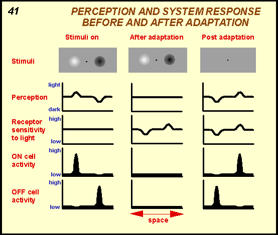
As fixation is maintained and the pigment molecules
reach a steady state, the ganglion cells gradually stop their discharge.
The result is that we no longer perceive the stimuli. Now when gaze is
shifted to the lower fixation spot, the uniform light energy emanating
from the left and right of the fixation spot impinges on the retina that
at the sites where the blobs had been have different ratios of isomerized
molecules compared to those that had been exposed to the background and
hence have different sensitivities. This means that the retinal region
where the dark blob had been became more sensitive to light and the region
where the light blob had been became less sensitive. As a result, the
uniform light that now impinges at these sites activates ON ganglion cells
where the dark blob had been and OFF cells where the light blob had been.
This in turn produces the percept of the negative afterimages.
|