
The Degen Lab
Nanoscale magnetic resonance imaging and spectroscopySpin physics and Nanomechanics
|
The Degen LabNanoscale magnetic resonance imaging and spectroscopySpin physics and Nanomechanics |
|
Techniques and
Instrumentation
|
  Basic
setup of a Magnetic
Resonance Force Microscope. An ultrasensitive silicon cantilever
carrying the sample is placed above a nanoscale magnetic tip. Spins in
the sample feel an attractive (or repulsive) magnetic force to the tip.
Rapid flipping of the spins by RF pulses drives the cantilever into
mechanical oscillation, which can be measured by Laser interferometry.
Three-dimensional scanning
of the cantilever over the magnetic tip then
produces a three-dimenional image of the sample's spin density. Basic
setup of a Magnetic
Resonance Force Microscope. An ultrasensitive silicon cantilever
carrying the sample is placed above a nanoscale magnetic tip. Spins in
the sample feel an attractive (or repulsive) magnetic force to the tip.
Rapid flipping of the spins by RF pulses drives the cantilever into
mechanical oscillation, which can be measured by Laser interferometry.
Three-dimensional scanning
of the cantilever over the magnetic tip then
produces a three-dimenional image of the sample's spin density. |
MRFM has enabled some remarkable experimental achievements over the last decade. These achievements include the detection of a single electron spin in 2004 [1] and the demonstration of nuclear magnetic resonance imaging of a nanoscale biological sample in 2009 [2,3]. Simultaneously, the technique has also contributed many concepts to nanoscale NMR spectroscopy and chemical contrast imaging.
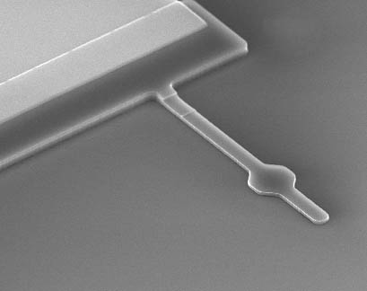 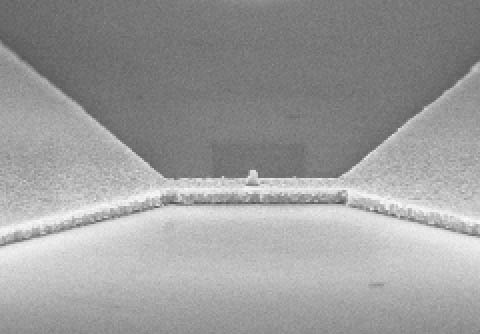 Ultrasensitive Si cantilever achieving Attonewton force sensitivity (left, length is 9 um); and nanoscale FeCo magnetic tip on top of an rf stripline (right, tip height is 250 nm). |
For our MRFM-related experiments we are currently assembling a custom-built, low temperature scanning force microscope that will enable highest sensitivity and have nanometer spatial resolution. The instrument combines a dedicated scanning force microscope with three-dimensional positioning functionality at sub-nanometer precision and stability. It will operate within a dilution refrigerator at temperatures between 100 mK and 4 K, as well as in magnetic fields up to 6 T. Key components to the setup will be lithographically-made silicon cantilevers and nanoscale magnetic tips, which we are currently fabricating.
Further reading:
[1]
D. Rugar, R. Budakian, H. J. Mamin, and
B. W. Chui, "Single spin detection by magnetic resonance force
microscopy", Nature 430, 329 (2004). (link)
[2] C. L. Degen, M. Poggio, H. J. Mamin, C. T. Rettner, and D. Rugar,
"Nanoscale magnetic resonance imaging", PNAS 106, 1313 (2009). (link)
[3] P. C. Hammel, "Imaging: Nanoscale MRI" (News & Views
article),
Nature 458, 844 (2009). (link)
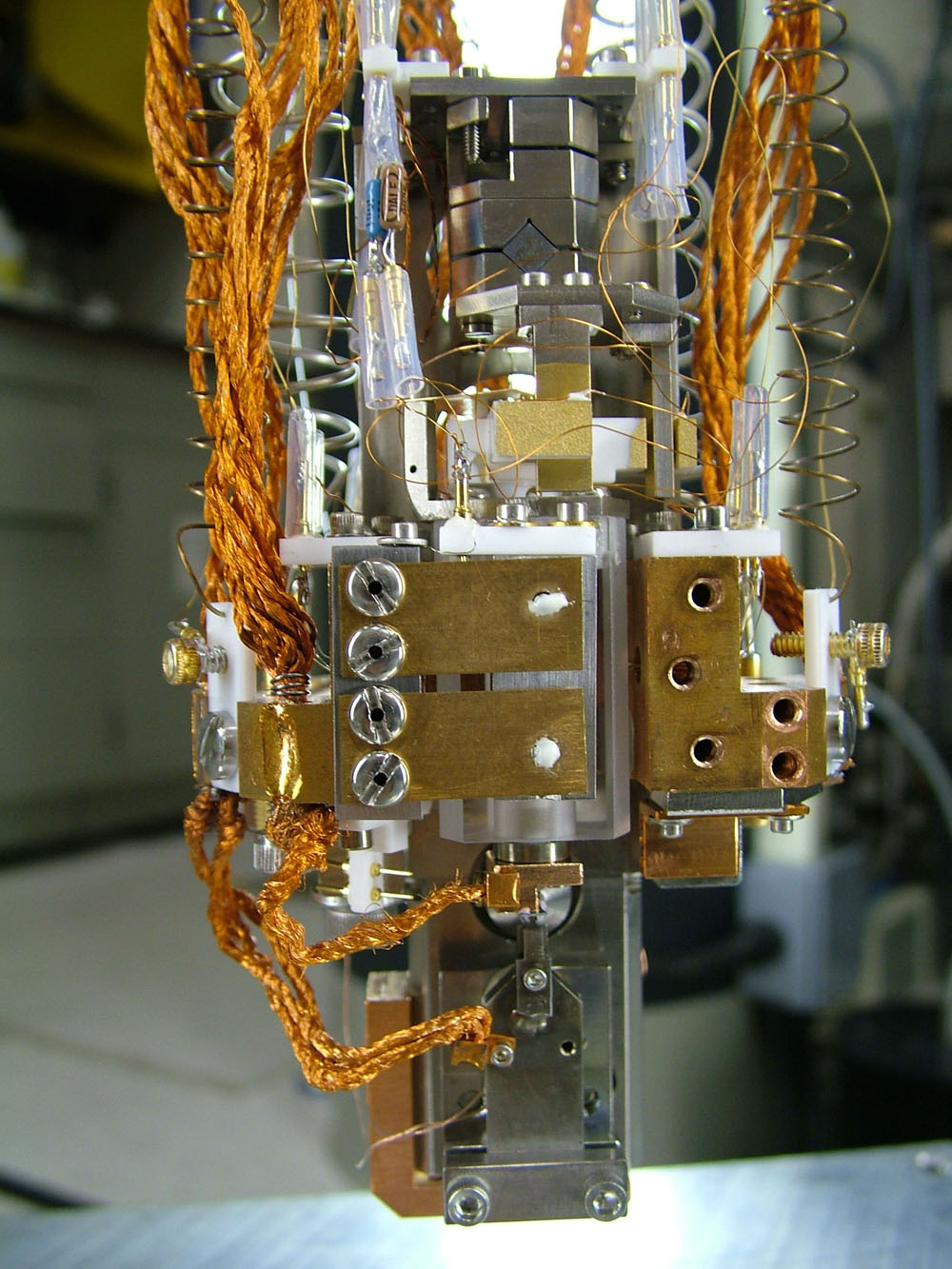 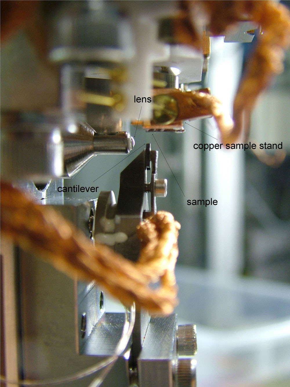 Front
and side views of the MRFM apparatus. Image
height is about 5 cm. Front
and side views of the MRFM apparatus. Image
height is about 5 cm. |
A disadvantage of MRFM is that it only performs well at cryogenic temperatures and in vacuum, which is not compatible with many biological (and other) applications. Therefore we are trying to come up with other ideas that might allow us doing the same experiments under ambient conditions.
In “scanning diamond magnetometry”, we use quantum optics to sense minute magnetic signals from samples via the fluorescent light emitted by a diamond defect (the so-called nitrogen-vacancy color center). Diamond defects carry a spin that is very sensitive to magnetic fields, owing to the Zeeman effect, and as the optical rate of emission depends on the defect’s spin state, we can measure small fields simply by looking at the fluorescence intensity [1].
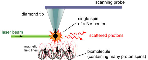 Basics of nanoscale magnetic microscopy using NV centers in diamond. |
Our goal is to place a diamond defect in a nanocrystal and attach to a scanning probe - in this way we can perform nanoscale imaging. Our scanning diamond magnetometer will have the capability to measure the dipole field of single electron spins, or monolayers of organic materials containing proton spins, at room temperature. Combination of diamond sensors with magnetic resonance spectroscopy will then allow us to perform nanometer-resolution chemical analysis of a wide variety of organic surfaces and other materials.
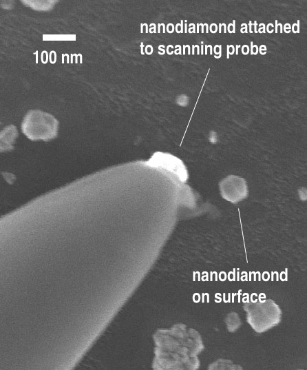  Left: Diamond nanocrystal attached to a tungsten STM tip Right: One-by-one positioning of seven nanodiamonds using a nanomanipulation stage in a scanning electron microscope (work together with Tjerk Oosterkamp and Ronald Hanson) |
For our diamond experiments we are currently assembling a dual confocal – atomic force microscope with optical access from both sides. The apparatus also incorporates a permanent magnet with precisely adjustable field orientation to access large bias fields for doing spectroscopy. This instrument will allow us to study individual diamond NV centers and to perform magnetic imaging and spectroscopy experiments on a range of transparent and opaque samples. A second important part of our experimental work is also to find efficient ways to fabricate, purify and chemically modify high quality diamond probes.
Further reading:
[1] C. L. Degen, "Scanning magnetic field microscope with a diamond single-spin sensor", Appl. Phys. Lett. 92, 243111 (2008). (link)