Digital Holographic Imaging of Microstructured and Biological Objects
Jose A. Dominguez-Caballero, Jerome H. Milgram, George Barbastathis
Sponsors: MIT Sea Grant
Digital Holographic Imaging (DHI) is a powerful technique that allows three-dimensional imaging by recording the optical wave field using a CCD array. This is followed by a numerical reconstruction of the image field. DHI is being employed to characterize microstructured and biological objects at relative far distances from the CCD with high lateral resolution. The amplitude and phase information can be subtracted from the reconstructed field, allowing us to obtain a more complete description of the sampled object.
The digital hologram is created by capturing the interference between the wavefronts scattered by an illuminated object and a reference beam. The intensity registered at the CCD plane is given by:

where r is the reference field and o is the Fresnel diffracted object field. The recorded intensity is then multiplied by the digitally generated version of the reference beam ( r*) in order to recover the virtual image. The next step is to reconstruct the object in the image plane by using the diffraction integral. The reconstruction is achieved using the convolution approach:

 is the reconstructed field in the image plane;
  and
and  are the forward and inverse Fourier Transforms respectively and h is the diffractive kernel. The reconstruction is optimized by using the FFT algorithms.
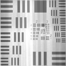
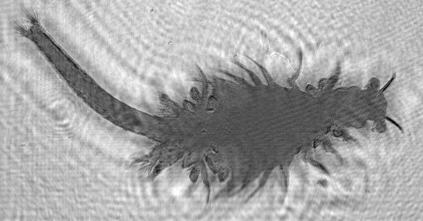
Experiments were done using an in-line configuration to reconstruct a USAF resolution target and a live brine shrimp. The recordings were made using a He-Ne Laser with  = 632.8nm. The beam was expanded and split into two paths of equal length forming a Mach-Zehnder Interferometer. The sampled object was placed in one of the paths forming the object beam. Neutral Density Filters were used to control the relative intensity between the object and reference beams. The hologram was captured using a CCD array of 4096x4096 pixels with a pixel size of 9µm and a fill factor of 100%. Figure 1 shows the reconstruction made for the USAF resolution target located 15mm apart from the CCD array. Figure 2 shows the reconstruction made for the live brine shrimp located at a distance of 167mm. The length of the brine shrimp is approximately 5mm.

Fig. 1. Reconstruction of a USAF resolution target: z = 15mm.

Fig. 2. Reconstruction of a live brine shrimp: z = 167mm.