The Neural
Control of Vision
B.
Wiring Diagram of the Visual System
 Figure
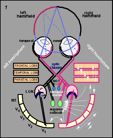
1 provides a schematic outline of the primate visual areas
and their connectivity. The retinal ganglion cells from the nasal half
of each retina send their axons to the contralateral half-brain whereas
the ganglion cells from the temporal hemiretinae project ipsilaterally.
As a result of this arrangement the left brain sees the right visual hemifield
and the right brain the left visual hemifield; the neural signals produced
by any one object in each eye end up in the same location in the brain. Figure
1 provides a schematic outline of the primate visual areas
and their connectivity. The retinal ganglion cells from the nasal half
of each retina send their axons to the contralateral half-brain whereas
the ganglion cells from the temporal hemiretinae project ipsilaterally.
As a result of this arrangement the left brain sees the right visual hemifield
and the right brain the left visual hemifield; the neural signals produced
by any one object in each eye end up in the same location in the brain.
The output cells of the retina are the retinal
ganglion cells. There are more than a million of them in each eye and
they connect to several brain structures that include the lateral geniculate
nucleus of the thalamus, the superior colliculus, nucleus of the optic
tract, and the terminal nuclei. The lateral geniculate nucleus (LGN) forms
the gateway to visual cortex where in primates most of the computations
for vision are carried out. The superior colliculus plays a central role
in eye-movement control which is discussed in the second section of this
web page.
In the retina, the LGN, and the visual cortex,
each neuron sees only a small portion of the visual field. This small
area is called the receptive field (RF) of the cell. The receptive fields
of retinal ganglion cells, LGN cells and primary visual cortex cells are
very small, often no more than a fraction of a degree in central vision.
As we ascend to higher levels in the cortex the receptive fields become
progressively larger. The visual field representation in most visual brain
structures is laid out in a neat, topographic order.
The circle shown that runs through the nodal point
of each eye and touches the point at which the two eyes fixate, is called
the Vieth-Muller circle or the horopter. Light originating from any object
that is positioned along this circle hits corresponding retinal points
in the two eyes from which the ganglion cells connect, via subcortical
structures, to corresponding brain regions. Stimuli that are either nearer
or further away activate non-corresponding retinal points.
|