The Neural
Control of Vision
C.
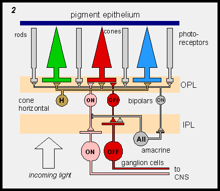
The Retina
 Five
major cell types have been identified in the retina: The photoreceptors
that form a single layer of cells against the inner wall of the eye, the
horizontal cells, the bipolar cells, the amacrine cells, and the ganglion
cells whose axons send the signals to the brain. There are two major classes
of photoreceptors, the rods and the cones. Rods are for night vision and
cones for day vision. Cones further subdivide into three color-selective
types in old world primates and in humans, that have peak sensitivity
in the short (blue), medium (green) and long (red) regions of the visible
spectrum. The horizontal, bipolar, amacrine and ganglion cells also come
in several subvarieties. Five
major cell types have been identified in the retina: The photoreceptors
that form a single layer of cells against the inner wall of the eye, the
horizontal cells, the bipolar cells, the amacrine cells, and the ganglion
cells whose axons send the signals to the brain. There are two major classes
of photoreceptors, the rods and the cones. Rods are for night vision and
cones for day vision. Cones further subdivide into three color-selective
types in old world primates and in humans, that have peak sensitivity
in the short (blue), medium (green) and long (red) regions of the visible
spectrum. The horizontal, bipolar, amacrine and ganglion cells also come
in several subvarieties.
It is convenient to think of retinal organization
as consisting of two major systems: the through system and the
lateral system. Starting with the photoreceptors, the through system
connects to the retinal ganglion cells via the bipolar cells. There are
two lateral systems that are produced by virtue of the horizontal cells
and the amacrine cells. These lateral networks give rise to the surround
organization of receptive fields as seen in the retinal ganglion cells
that will be discussed shortly. The interconnections among receptors,
horizontal cells and bipolar cells take place in the outer plexiform layers
(OPL); interconnections between bipolar cells, amacrine cells and retinal
ganglion cells take place in the inner plexiform layers (IPL).
The rods and cones converge on retinal ganglion
cells via different pathways. The cones connect with ganglion cells via
cone bipolar cells. The rods connect with the ganglion cells via rod bipolars
and amacrine cells.
The information from the retina is sent to the
brain by the axons of the retinal ganglion cells. There are several different
classes of retinal ganglion cells that differ not only in their response
characteristics, but also are distinct anatomically. The two classes shown
in Figure 2 are the ON and OFF which
will be discussed below.
|