The Neural
Control of Vision
D.
The Lateral Geniculate Nucleus of the Thalamus
 Figure
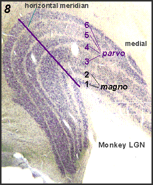
8 shows a coronal section through the lateral geniculate
nucleus (LGN) of the rhesus monkey. This thalamic structure receives profuse
projections from retinal ganglion cells. In central retina (up to about
17 degrees of eccentricity), this structure has 6 layers. The number of
layers representing larger eccentricities reduces to 4. The individual
layers are segregated according to left and right eye input and also according
to cell types. Layers 6, 4 and 1 receive input from the contralateral
eye, the rest from the ipsilateral eye. The upper four parvocellular layers
receive input from the retinal midget ganglion cells whereas the lower
two magnocellular layers that have larger cells, receive input from the
retinal parasol ganglion cells. The layout in the LGN is orderly; the
horizontal meridian is along the line shown, with the medial portion of
the LGN representing the lower, and the lateral portion the upper visual
field. Antero-posteriorly in this structure we move from peripheral to
central representation of the visual field. In each LGN the contralateral
visual hemifield is represented as already shown in Figure 1. The interlaminar
layers of the LGN have very small cells of a different type that also
receive input from the retina; at this time little is known about this
rather heterogeneous group of cells. The receptive field properties of
cells in the parvocellular layers are similar to those of the midget ganglion
cells; the receptive field properties of the magnocellular layers similar
to those of the parasol cells. Figure
8 shows a coronal section through the lateral geniculate
nucleus (LGN) of the rhesus monkey. This thalamic structure receives profuse
projections from retinal ganglion cells. In central retina (up to about
17 degrees of eccentricity), this structure has 6 layers. The number of
layers representing larger eccentricities reduces to 4. The individual
layers are segregated according to left and right eye input and also according
to cell types. Layers 6, 4 and 1 receive input from the contralateral
eye, the rest from the ipsilateral eye. The upper four parvocellular layers
receive input from the retinal midget ganglion cells whereas the lower
two magnocellular layers that have larger cells, receive input from the
retinal parasol ganglion cells. The layout in the LGN is orderly; the
horizontal meridian is along the line shown, with the medial portion of
the LGN representing the lower, and the lateral portion the upper visual
field. Antero-posteriorly in this structure we move from peripheral to
central representation of the visual field. In each LGN the contralateral
visual hemifield is represented as already shown in Figure 1. The interlaminar
layers of the LGN have very small cells of a different type that also
receive input from the retina; at this time little is known about this
rather heterogeneous group of cells. The receptive field properties of
cells in the parvocellular layers are similar to those of the midget ganglion
cells; the receptive field properties of the magnocellular layers similar
to those of the parasol cells.
|