The Neural
Control of Vision
H. The Function of the Midget and Parasol Channels in Vision
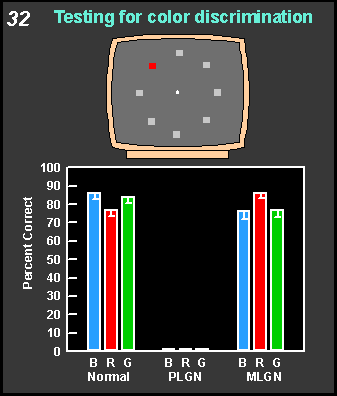
 The
upper part of Figure 32 shows how
color vision is tested. One of the targets is of a different color than
the other in this discrimination task. The graph below shows a monkey's
performance when the animal had to discriminate a red, blue or green stimulus
from white ones. The monkey does well at intact and magnocellular lesion
sites (MLGN), but cannot discriminate colors at the parvocellular lesion
site (PLGN). The
upper part of Figure 32 shows how
color vision is tested. One of the targets is of a different color than
the other in this discrimination task. The graph below shows a monkey's
performance when the animal had to discriminate a red, blue or green stimulus
from white ones. The monkey does well at intact and magnocellular lesion
sites (MLGN), but cannot discriminate colors at the parvocellular lesion
site (PLGN).
|