What is MEG?
Basic Principles of Magnetoencephalography
(click on any figure to enlarge)
Content from www.4dneuroimaging.com
© 4-D Neuroimaging
|
What is MEG? |
Basic Principles of Magnetoencephalography (click on any figure to enlarge) Content from www.4dneuroimaging.com © 4-D Neuroimaging |
|
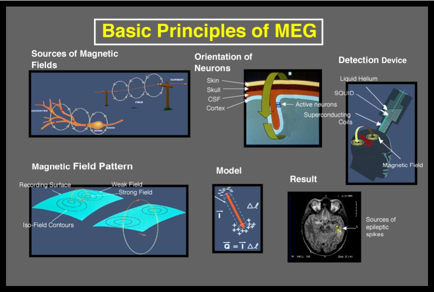
Magnetoencephalography (MEG) is a non-invasive neurophysiological technique that measures the magnetic fields generated by neuronal activity of the brain (Figure 1). The spatial distributions of the magnetic fields are analyzed to localize the sources of the activity within the brain, and the locations of the sources are superimposed on anatomical images, such as MRI, to provide information about both the structure and function of the brain (Figure 2). The principle features
of MEG are: Magnetic fields are found whenever there is a current flow, whether in a wire or a neuronal element, as illustrated in the upper left panel of Figure 3. The magnetic field passes unaffected through brain tissue and the skull, so it can be recorded outside the head (upper middle). The magnetic field is extremely small, but can be detected by sophisticated sensors that are based on superconductivity (upper right). By analyzing the spatial distributions of magnetic
fields (lower left), it is possible, by using a model such as a single
equivalent current dipole (lower middle), to estimate the intracranial
localization of the generator source and superimpose it on an MRI (lower
right). The spatial and temporal properties of MEG are
illustrated in the left panel of Figure 5. Only MEG has extremely high
temporal and spatial resolution, as represented in the lower left section
of the graph. Other functional modalities, except invasive EEG (iEEG),
have either poor temporal or spatial resolution. Clearly iEEG has the
distinct disadvantage of being invasive. MEG is a unique and effective diagnostic tool for evaluating brain function in a variety of surgical planning applications. The fundamental advantages of MEG are listed in Figure 6. |
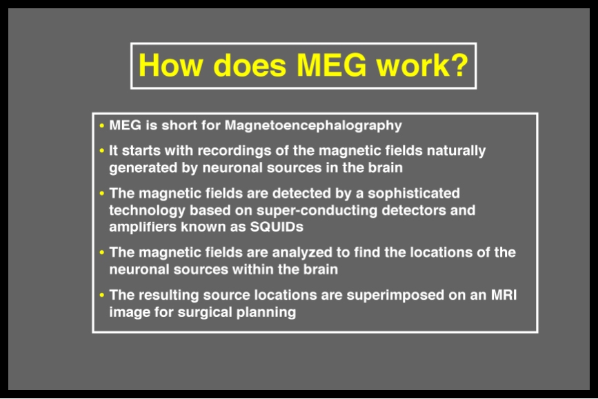
Figure 1. Basic principles of MEG.
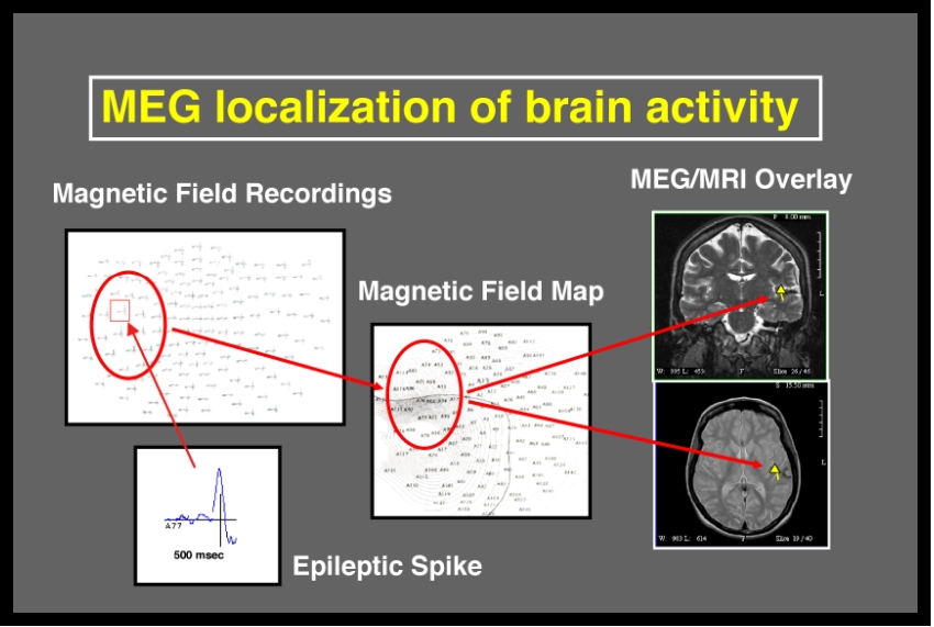
Figure 2. MEG combines functional information from magnetic field recordings with structural information from MRI.
Figure 3. Electrical activity in neurons produces magnetic fields that can be recorded outside the skull and used to calculate the locations of the activity within the brain.
Figure 4. The sequence of steps to localize sources of neuronal activity from time-domain recordings to MRI overlay.
Figure 5. MEG has the advantages of very high temporal and spatial resolution, however it requires highly sensitive instrumentation and sophisticated methods for eliminating environmental magnetic interference.
Figure 6. Advantages of MEG for surgical planning. |
|
What is MEG? • About Our System • People • Being a Subject • Getting Here • More Info |