Laboratory of Molecular Architecture
Molecular Machine Group, Media Lab


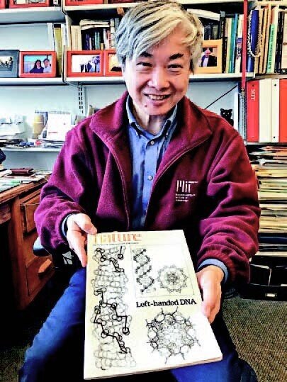
Shuguang Zhang
"If you ask big questions, you get big answers." --Francis Crick
Latest News

MIT News (July 21, 2023)
New sensor mimics cell membrane functions | MIT News | Massachusetts Institute of Technology
An MIT-led team designed a sensor that may be deployed to screen patients for hard-to-diagnose cancers, or metastatic tumors. The device draws inspiration from the membrane that surrounds all cells.
Zhang, S. (2022) Life has its ups and downs, always ask questions. Molecular Frontiers Journal, Vol. 6, No. 01n02. [local pdf] A brief autobiography.


Proteins may halt the severe cytokine storms seen in Covid-19 patients
MIT News, April 16, 2020.
Water-soluble cytokine receptors fused with Fc domain of IgG may be therapeutic for cytokine storms
Shuguang Zhang receives Emil Thomas Kaiser Award
MIT Media Lab, March 13, 2020
The Protein Society announces its 2020 award recipients
The Protein Society, AAAS, March 12, 2020
Dr. Zhang is widely seen as a founder of the field of peptide nanomaterials. He discovered a class of ionic self-complementary peptides that undergo molecular self-assembly to form well-ordered nanofibers and membranous structures.

Making chimeric versions of chemokine receptors.
Chemical and Engineering News, December 1, 2019, Volume 97, Issue 47.
QTY code designed thermostable and water-soluble chimeric chemokine receptors with tunable ligand affinity. [link]
Proceedings of the National Academy of Sciences, November 27, 2019

We Are All Chinese Scientists
‘Psychological fear’: MIT scientists of Chinese origin protest toxic US climate
Researchers describe how a government crackdown on foreign influence is affecting them following a statement of support from their university.
Nature News, July 2, 2019
China Science Communication interview (in Chinese)
Shanghai, November 7, 2018
The Excitement of Discovery: Selected Papers of Alexander Rich. A Tribute to Alexander Rich. [link] https://doi.org/10.1142/11055 | January 2019. Pages: 624. Edited By: Shuguang Zhang (Massachusetts Institute of Technology, USA)
Curiosity-driven research: Fractals and QTY Code; a talk for high school students given at the 2018 Molecular Frontiers Symposium held on November 16-17, 2018 at the MIT Media Lab, Cambridge, Massachusetts.
Scientists alter membrane proteins to make them easier to study. (Centre for Structural Systems Biology, 8/29/2018) [link]
Following a code for swapping amino acids makes membrane proteins water soluble. (Chemical & Engineering News, 8/29/2018) [link] [local pdf]
Scientists alter membrane proteins to make them easier to study (ScienceDaily, 8/28/2018) [link] [local pdf] (MIT News, 8/27/2018) [link] [local pdf]
Research Highlights, 1967-2016. (MIT Office of the Provost, Institutional Research)
[link] [local pdf]
Growing a business from the lab (MIT News, 02/03/2014)
[publisher link] [local pdf]
Chinese investors tap US Biotechs (Nature Biotechnology Interview, 02/2013)
[local pdf]
Harnessing nature’s solar cells (MIT News, 2/3/2012) [local pdf]