
Scientists Join Forces in Search for Faster Detection of Harmful Algal Blooms
by Tracey Crago, WHOI Sea GrantFor Don Anderson, a senior scientist at the Woods Hole Oceanographic Institution (WHOI), scientific meetings offer a chance to catch up with colleagues and hear about their latest work. But at the 2001 satellite workshop to the Gordon Conference on Mycotoxins and Phycotoxins, it was a foray into a session unrelated to his own work that proved especially fruitful.
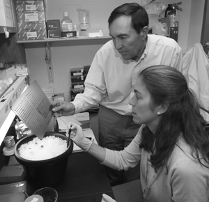
Deana Erdner and Don Anderson discuss the results from some of their lab-based experiments on HAB species.
Jason Epstein, then a post-doc in David Walt's lab at Tufts University in Medford, was giving a presentation on the "optical nose," an application using multi-bead arrays on the ends of optical fiber bundles to "sniff out chemicals" in the environment. The talk intrigued Anderson, one of the world's leading experts on harmful algal blooms (HABs), whose lab develops molecular probes for a number of HAB species. Curiosity piqued, Anderson skipped the next session, found the post-doc, and they sat down and talked for several hours.
Soon, Anderson and Epstein came up with a few ideas for experimentation. Back at WHOI, Anderson and his lab members faced the first challenge: "how can we get our probes onto the ends of their fiber bundles and make them work?" Results from a few "quick experiments," says Anderson, led to Sea Grant funding through a National Strategic Investment award to develop an optical fiber-based technology for HAB cell detection.
Blooms of certain HAB species can cause mortality in fish, toxicity in shellfish, and problems even higher up the food chain, in marine mammals and humans. Less publicized are the ensuant economic effects associated with HABs, estimated by WHOI marine economist Porter Hoagland at $82 million last year in the U.S. alone (Hoagland and Scatasta, 2005, in press). That figure accounts for costs in categories including public health, commercial fisheries, recreation and tourism, and monitoring and management. Last spring, a particularly large bloom of Alexandrium fundyense (the dinoflagellate that produces saxitoxins, responsible for causing paralytic shellfish poisoning) ranging from the Gulf of Maine to Cape Cod and into Buzzards Bay, was responsible for the closures of 1.3 million acres of state shellfish beds. For weeks, recreational and commercial shellfishing along most of the state's coastline were shut down, as were aquaculture operations.
HAB monitoring programsóonce focused on the detection of algal toxins in shellfishónow include monitoring of the plankton for HAB species. While this represents progress, the sheer number of samples collected is rising sharply each year, as are the processing times and expense. What's needed, says Anderson, is innovation. One possible solution: automated instruments that can detect HAB species remotely. The Anderson-Walt collaboration is a first step in that direction.
One of the first considerations faced by the two labs was the use of fiber optic technology with a biological organism. Walt's previous work involved detection of chemicals or biological molecules using direct hybridization. Hybridization is a process in which two complementary strands of DNA (or one each of DNA and RNA) are joined to form a double-stranded molecule. Anderson's HAB cell cultures would require the use of a two-step, or "sandwich" hybridization process: using two probes specific to large subunit ribosomal RNA (rRNA) in the target HAB organism.
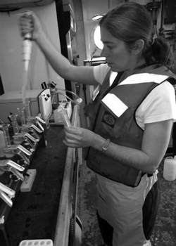
Deana Erdner filters seawater samples containing the dinoflagellate Alexandrium fundyense aboard the R/V Oceanus during a HABs cruise in Massachusetts Bay last spring. A. fundyense produces a toxin responsible for causing paralytic shellfish poisoning, or PSP.
The fiber optic technology being explored by Anderson and Walt involves the use of thousands of imaging optical fibers held together in a millimeter-sized bundle. Etched into the end of each optical fiber (3-7 microns (µm) in diameter) is a tiny well or pit. A microsphere, or bead, fits into the pit. The beads are coated with different concentrations of fluorescent dye, then, in this case, coupled with a specific HAB probe, known as the capture probe. This probeódeveloped by the Anderson lab with partial funding from Sea Grantómatches a specific region of the rRNA sequence in the target HAB species. Once the beads have been coded and coupled with the specific probes, they are loaded onto the end of a fiber optic bundleóup to 50,000 fibersóto form an array. Each array may contain multiple types of beads, says Deana Erdner, a research associate in Anderson's lab. This makes it possible, she says, to detect multiple HAB species at once, a definite advantage to the technology.
Scientists take a picture of the array's fluorescence, which corresponds with the concentration levels of the dye in the beads. "That picture," says Erdner, "gives us a map of where every bead type is on a specific bundle."
The prepared optical fiber bundle is then dipped into a plankton lysateówhat Erdner calls "cell soup." It's actually a solution containing the nucleic acids from ruptured plankton cells, including some from the target HAB organism. After a few minutes, the first hybridization occurs, says Erdner, and the RNA from the HAB cells "matches up and sticks to" only those beads containing the capture probe specific to the species of interest.
A microscope-based imaging system developed by Walt is used to capture light from the second hybridization, in which a fluorescently-labeled "signal" probe attaches to a second binding site on the HAB rRNA. The "sandwich" on each bead is now complete: signal probe-target rRNA-capture probe. The resulting fluorescent signal from each bead (which is coated with thousands of capture probes) gives "a direct measure of how many molecules are bound to the bead," says Erdner. Using computer imaging software programmed to account for background fluorescence, detection limits, and standard deviation, this image can be decoded by comparing it to a control image taken earlier in the process.
The results have been successfulóand consistent. "We have shown that the technique works with all three of our target HAB species," says Erdner. Those species, Alexandrium fundyense, Alexandrium ostenfeldii (a toxic dinoflagellate that produces spirolides, a family of neurotoxins), and Pseudo-nitzschia (a toxic diatom that produces domoic acid, the causative agent of amnesic shellfish poisoning), are all found in the Gulf of Maine.
What's more, says Erdner, they have been able to refine the detection limit for the single-probe arrays for both A. fundyense and P. australis to 5 cells/sampleófar better than the original goal of 50 cells/sample. A new single-probe array for A. ostenfeldii has been designed and the team is working to define its detection limit.
Next up, says Erdner, is the automation piece. "With most of the lab characterization done, we're ready to try a field-based system, ground-truthing [the process] with seawater samples." This advance would be welcome news for those monitoring HABs worldwide, not to mention local officials. In last year's record-breaking outbreak of A. fundyense, samples were collected weekly along nearly 80 percent of the state's waters, including areas that had never before experienced outbreaks of the toxic species. Though Walt's instrument is not yet ready for deployment in a harsh ocean environment, Erdner hopes it will be ready within 5-10 years.
"There is a lot of instrumentation in development out there that could help push along the automation of this technology," says Erdner. She points to work underway by colleagues at WHOI (Heidi Sosik and Rob Olson, who have developed Imaging FlowCytobot, an automated underwater microscope that counts and identifies phytoplankton), and those at Monterey Bay Aquarium Research Institute (Chris Scholin, Anderson's former student, who has developed the Environmental Sample Processor, or ESP, an instrument that collects water samples and automates the application of molecular probes to identify marine planktonic organisms). Both, she says, may represent possible interfaces for the technology that the Anderson and Walt laboratories have developed.