|
|
Bone marrow stromal cells (BMSCs) include a subset of stem cells that are considered promising for developmental studies and therapeutic applications. In this work led by BioSyM PI Prof.Krystyn Van Vliet, it has been observed that BMSC colony formation in real time via time lapsed optical imaging and analysis, to quantify whether and how heterogeneity emerged over multiple cell divisions spanning the duration of a typical colony formation unit assay. These analyses demonstrate that such colonies are neither homogeneous subpopulations of stem cells nor necessarily derived from single originating cells. These direct quantitative observations and visualizations of colony formation provide new insights that are motivated by significant implications for both basic research and stem cell-based therapies. |
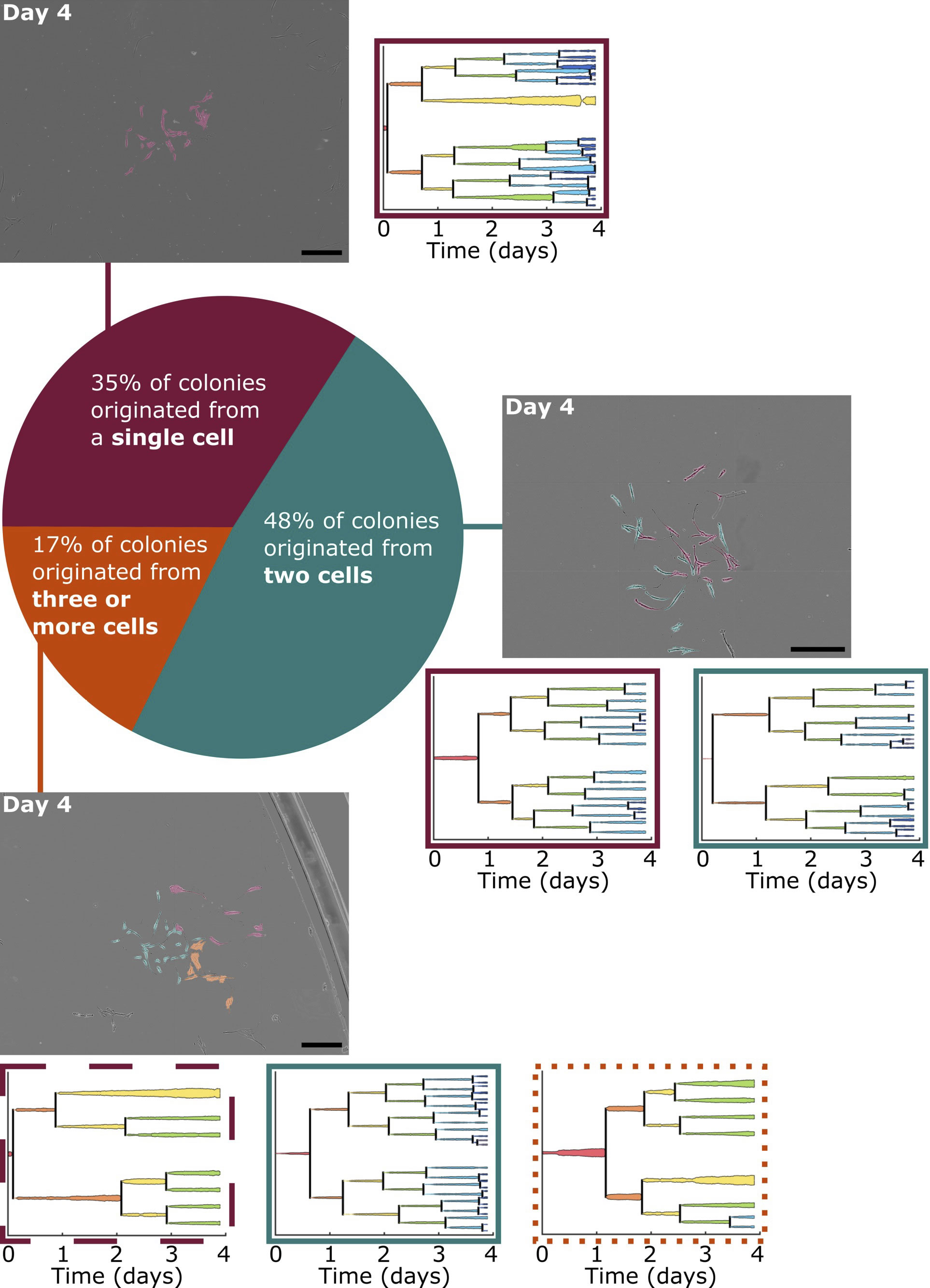 |
Colonies observed after four days of growth varied by the number of originating progenies. Of all locations in which colony formation was observed, approximately 35% originated from a single tracked cell, 48% originated from two analyzed cells, and the remainder formed from three or four originating cells that attached near each other at the start of the experiment. Phase contrast images of three representative colonies after four days of growth are displayed, organized by the number of analyzed cells the colonies originated from. Images are montages of nine or sixteen fields of view and were altered post-experiment to artificially color the BMSCs by the progeny they belong to. Cells without color added are BMSCs that migrated into the montaged field of view after initial imaging. Lineage trees of the colonies’ originating cells are also presented, where the width of the lineage lines is representative of the respective cell spread area at each 15 min time point. Lineage trees and images of all colonies at several time points are presented in S1 Fig. Lineage tree outlines indicate the proliferative capacity of the progeny. Dashed line (- -): progeny classified as slow proliferator; dotted line (. . .): progeny classified as moderate proliferator; solid line (–): progeny classified as fast proliferator. Scale bars = 0.5 mm. |
......More BioSyM Highlights |
|
Recent Publications
- High-resolution Imaging of Nuclear Dynamics in Live Cells under Uniaxial Tensile Strain , Journal of Visualized Experiments
- Emergent heterogeneity in putative mesenchymal stem cell colonies: Single-cell time lapsed analysis, PLOS One
- “Phthalimide Derivative Shows Anti-Angiogenic Activity in a 3D Microfluidic Model and no Teratogenicity in Zebrafish Embryos”, Frontiers of Pharmacology
- A Robotic Microscope System to Examine T Cell Receptor Acuity Against Tumor Neoantigens: A New Tool for Cancer Immunotherapy Research, IEEE Robotics and Automation Letters.
- "Engineering silver-zwitterionic composite nanofiber membrane for bacterial fouling resistance" , J. Applied Polymer Science,
- "Calcium-mediated Protein Folding and Stabilization of Salmonella Biofilm-associated Protein A", Journal of Molecular Biology
- "A combined microfluidic-transcriptomic approach to characterize
the extravasation potential of cancer cells", Oncotarget
- "Improving hematopoietic recovery through modeling and modulation of the mesenchymal stromal cell secretome", Stem Cell Research & Therapy
- "Quantitative phase microscopy of red blood cells during planner trapping and propulsion", Lab on a Chip
Meet the Principal Investigators, Collaborators, researchers, students and staff of SMART-BioSyM
Read about our research thrusts/projects, lab facilities and publications
|