What are the structural mechanisms that underlie modularity in the brain? My research attempts to answer this question by using structural connectivity as measured by diffusion weighted imaging (DWI) in order to predict the boundaries of regions that are specialized for particular behavioral or neural functions. These boundaries delineate either functionally defined regions through the use of fMRI, or anatomically defined regions not yet discernible with common structural MRI. I argue that in either case, these regions are fundamentally specialized due to their characteristic connectivity fingerprints.
My research program also focuses on the relationship between connectivity and plasticity, especially through development. The brain is adaptive, and it undergoes functional changes as we mature and learn new skills. Experience and expertise are known to modulate the functional properties of specialized brain regions, and these functional changes should coincide with distinct changes in connectivity patterns. I study these changes in a variety of contexts, including typical and atypical development, as well as how they relate to individual differences that predict treatment outcomes in pathology.
My research program also focuses on the relationship between connectivity and plasticity, especially through development. The brain is adaptive, and it undergoes functional changes as we mature and learn new skills. Experience and expertise are known to modulate the functional properties of specialized brain regions, and these functional changes should coincide with distinct changes in connectivity patterns. I study these changes in a variety of contexts, including typical and atypical development, as well as how they relate to individual differences that predict treatment outcomes in pathology.
Predicting neural responses from connectivity patterns
Function predicted by structural connectivity
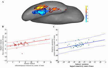
I'm interested in the anatomical underpinnings of brain function. Specifically, does there exist a connectivity fingerprint that delineates a functionally distinct region from its neighbors?
I first turned to the fusiform gyrus as a testing ground for this question (Saygin et al. 2012), because this region contains a highly selective face area. Specifically, I tested whether structural connectivity to each brain region, as measured with DWI, can accurately predict the location and degree of face-selectivity in the fusiform gyrus. Such an approach would strongly demonstrate a relationship between gradients of face-selectivity and connectivity. The results showed that it was indeed possible to accurately predict functional activity to faces in this brain region and that these predictions outperformed two control models and a standard group-average benchmark in two separate groups of participants. Additionally, this approach identified cortical regions whose connectivity was highly influential in predicting face selectivity within the fusiform, suggesting a possible mechanistic architecture underlying face processing in humans.
I'm extending this research method to 1) other brain regions (Osher et al. 2015) and 2) other domains (scene, body, and object perception, Theory of Mind processing). I'm also investigating the limits of the current approach and comparing standard diffusion imaging and tractographic methods to the new, one-of-a-kind technology offered by the Connectome scanner (below).
I also use tractography to compare the connectivity patterns of cortical regions that possess highly similar or distinct functions. For example, regions that are involved in language processing may be adjacent to regions recruited during high cognitive demand; we hypothesize that, although these regions are adjacent to one another, they may be more strongly connected to other regions with similar functional properties than to each other.
Connectome imaging and validation
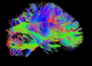
I'm using the new Connectome scanner at MGH (part of the Human Connectome Project), a one-of-a-kind, new technology for diffusion imaging that offers a substantial increase in signal quality and hence may afford significant improvements in tractography. In this project, I'm defining about 20 well-established regions with distinct functional profiles, and using new tractographic methods to discover the anatomical connections of each functionally-defined region to the rest of the brain. I'm also comparing standard diffusion imaging to the Connectome imaging in an effort to better understand the benefits of this new technology.
Structure-function relationships in development and disorders
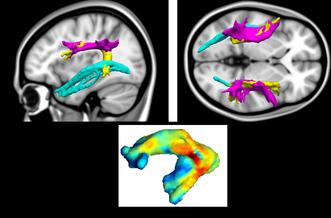
I'm currently investigating the development of the anatomical foundations of brain function. I'm interested in how functionally-specific brain regions become so specific and why the immature brain is more plastic or more amenable to acquiring different functions with brain injury. Some of my current investigations include:
- identifying children at risk for dyslexia based on tract integrity (figure on left; Saygin et al. 2013)
- characterizing differences between structural connectivity predictors of brain function in typical children vs. adults and ASD children vs. typical children
- exploring connectivity patterns that define cortical plasticity in congenitally blind children and adults
- determining predictors of treatment outcome for Social Anxiety Disorder (Doehrmann et al. 2012, Manning et al. 2015, Whitfield-Gabrieli et al. 2015)
Predicting anatomical boundaries from structural connectivity
Structural connectivity to define anatomical boundaries
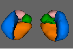
I use tractography and connectivity patterns derived from animal studies to create finer distinctions in the human brain than what is currently available through standard MRI. Finer anatomical distinctions will allow us to better constrain functional regions of interest and to provide more rigorous foundations for network analyses. I've used this approach (called TractSeg) to segment the various amygdalar subnuclei (Saygin et al. 2011). The figure on the left is a 3-D rendition of an individual participant's right and left amygdala, segmented into four major subdivisions based on tractography.
Development of structural connectivity
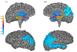
Tractographic segmentation can lead to interesting results. It turns out that while TractSeg results in consistent subnucleus definitions in adults, in children, the tractographic connectivity patterns are highly variable (Saygin et al. 2015). The connection patterns of the amygdala with certain brain regions are stronger in children than in adults (as shown on the left in blue colors). Other regions (yellow colors) grow increasingly connected to amygdala through development.
I'm currently investigating how the healthy development of the amygdala's connection patterns compare to those in ASD. It will be interesting if the connectivity differences are related to the functional differences that are usually reported in studies of ASD, such as the lower functional responses to faces in the fusiform.
I'm currently investigating how the healthy development of the amygdala's connection patterns compare to those in ASD. It will be interesting if the connectivity differences are related to the functional differences that are usually reported in studies of ASD, such as the lower functional responses to faces in the fusiform.
Validation through high-resolution structural MRI and histology
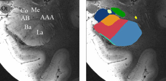
To validate the segmentation of amygdala nuclei, I'm using ultrahigh-resolution imaging of ex-vivo (post-mortem) brain samples on a 7T magnet (Saygin et al. in preparation). This gives us great resolution to visualize even finer boundaries. We can manually segment these images into 10 nuclei and validate the labeling through histology. I then hope to use diffusion weighted imaging and probabilistic tractogrpahy to explore the connectivity patterns of the nuclei and compare these to the patterns of connectivity that we used in TractSeg which were based off of animal studies.