Introduction
Definition of Life
Motivation
Preliminary
Steps
Present Life
Past Life
Geological Survey
Sample Collection
Spectroscopic Analysis
Organic Analysis
Biology Experiments
Thin Section
Isotope Analysis
Experimental Design
Motivation for Thin Section Analysis
When we look for fossils on Mars, we want to try to
emulate the process used on Earth to look for fossils as much as possible.
The process on Earth begins with a general geological survey to
identify potential fossil-bearing rocks—our astronauts will do this, and
the details are described in the section of this report on the geological
survey. The next step on
Earth is a visual analysis of the rock to look for cellular fossils, which
are omnipresent in many kinds of Earth rocks.
If a rock on Earth formed under conditions favorable to life, and
has not been too heavily altered by the ravages of time, it is bound to
contain fossils. Due to this
constant presence, a number of techniques have been developed to analyze
these fossils in various levels of detail.
The problem, however, is identifying which of the following options
is best suited to use on another planet, where the very presence of
fossils is questionable.
Options
Thin Section Inspection: One of the
most common techniques used to identify fossils on Earth is the
preparation and inspection of a thin section.
A thin section is essentially a very thin slice of rock mounted on
a microscope slide. The slice
is so thin as to be translucent, and highly polished.
It can be examined under a microscope to look for morphological
fossils—i.e. the shapes of the remains of long-dead organisms.
By adding a polarizing filter to the microscope, a thin section can
also be used to characterize the types of minerals present in a rock,
information useful in identifying whether the rock formed under conditions
favorable to life. This
method, however, usually requires a long and tedious series of polishing
steps.
Chemical
Fossil Extraction: Another way to look at microscopic fossils is
to dissolve the surrounding rock away, leaving only the fossils. The techniques to do this are well established, and allow
microscope examination of fossils in somewhat greater detail than in thin
section. However, it has a
number of drawbacks. It
requires the use of several extremely dangerous chemicals, including
highly corrosive hydrofluoric and nitric acids.
It also requires knowledge of the composition of the rock, in order
to know which chemicals to use to dissolve the rock.
This knowledge is often obtained from a thin section.
Finally, it requires the use of large amounts of water, which will
be a precious resource on Mars. (Brasier, 1980)
Electron
Microscope Inspection: An electron microscope can examine fossils in
much greater detail than any method using a light microscope.
Traditionally, however, electron microscopes have required involved
sample preparation, which can introduce artifacts that look like fossils.
While new types of electron microscopes are now available that
require no sample preparation, other problems remain.
Electron microscopes are heavy and power-hungry.
They use very sensitive detectors, which could easily be corrupted
by radiation in space. Finally,
they examine specimens in such great detail that their field of view is
very restricted, making it difficult to find fossils to examine.
Choice
From the above options, it is clear that the analysis of a thin section is the best way to visually look for fossils. Automating the process can eliminate its tedium. Machines exist which can prepare thin sections with no human intervention, and one will be used on the mission to prepare thin sections.
Principles of operation
Thin sections will be prepared using an automatic
thin-section-preparing device manufactured by Microtec Engineering, Inc.
The particular model we will use can go from a rough chunk of rock
to a finished slide with no human intervention, all in relatively little
mass and power (Microtec, 2000). Since it is a mechanical device, however, we will bring
enough spare parts to replace everything that could possibly break or wear
out in the machine.
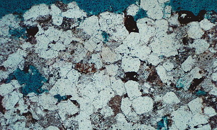
The thin sections produced will all be analyzed under a binocular polarizing microscope to determine whether they meet the criteria for fossil life, presented earlier in this report.. Fossils should appear as brownish structures in the thin section due to their high carbon content (Schopf, 1999). The polarizing filter will be used on each section to identify the types of minerals present in the sample. Different minerals behave differently under polarized light, and so can be identified from a chart after observing their behavior. For fossil-bearing rocks, the mineral composition can indicate some of the rock’s formation conditions, and thus determine whether the sample meets another condition for fossil life.
Equipment
|
Item |
Cost |
Mass |
Power |
|
thin sectioner |
~$10,000,000 |
149 kg |
550 W |
|
spare parts |
~$1,000,000 |
~150 kg |
N/A |
|
polarizing microscope (2) |
~$10,000 |
~4 kg each |
~20 W each |
|
slides (~1000) |
~$1,000 |
~10 kg |
N/A |
|
digital camera |
~$10,000 |
~1 kg |
~5 W |
|
lens cleaning tissue |
insignificant |
~1 kg |
N/A |
|
slide storage boxes |
~$100 |
~5 kg |
N/A |
|
hand lens (4) |
~$100 |
~2 kg |
N/A |
| distilled
water |
N/A |
~50 kg |
N/A |
Protocol
- Examine
the rock specimen with a hand lens to determine if it contains any
fine layers. Such
layering could suggest that the rock was built by microorganisms, in
which case fossils would be located between the layers.
Enter into the computer the specimen number, slide number, and
a brief description of the sample.
If the sample is layered, allocate two slides to it, and
indicate that one is parallel to the layers and one is perpendicular.
- If
the rock lacks layers, simply use the rock saw on the thin sectioner
to slice off a fragment approximately two inches square. If it has layers, prepare one piece cut parallel to the
layers and one perpendicular, taking care that the pieces remain
matched with the appropriate slides.
- Insert
the rock piece and slide into the thin sectioner.
It should be ready at a thickness of about 30 microns,
depending on the type of rock. Retrieve the prepared slide from the device.
- Examine
the slide at low magnification under a binocular microscope, so that
the entire slide is visible. Focus
the microscope appropriately, and replace the eyepieces with the
digital camera. Take one
picture of the section as a whole, and then replace the eyepieces.
- Examine
the slide at 500x magnification.
Be sure to cover the entire area of the slide, and note any
potentially fossil-like structures.
Upon encountering any such structure, take a digital picture.
- Upon
completion of examination, note the presence of any of the following
features: filaments or
clusters, large numbers of similar structures, variations between
structures similar to that produced by genetic variation, dividing
cells, brownish color
- Examine
the slide under high magnification on the polarizing microscope.
Determine the mineral phases present, and take at least one
picture of a representative sample of grain distributions, sizes,
shapes, and types.
- If
the structures in the rock meet all five of the above criteria, and/or
are in a type of rock that formed under conditions favorable to life
(sandstone, shale, mudstone, fine-grained silica, water-soluble
minerals), mark the slide as high priority and file it away.
Otherwise, mark the slide as low priority and file it away.
In addition, return to sites that yield high priority samples
to obtain new samples for confirmation.
- Regularly
filter the water in the thin sectioner, and replace it if it gets too
dirty or too low.
Use of this experiment
This experiment shall be conducted on any sample of
rock that has been returned from the field.
If field analysis, including naked eye inspection and alpha proton
x-ray spectrometer results, suggests that the rock will not contain
fossils, analysis can be restricted to the polarizing filter to determine
its mineral composition for geological interest.
Time for experiment
1 hour
References
Cradle
of Life: the Discovery of Earth’s Earliest Fossils. Schopf, J. William. 1999.
Princeton, NJ: Princeton U. Press.
Microfossils.
Brasier, M. D. 1980. London:
Chapman & Hall.
Microtec Engineering corporate web site: http://www.microtec.gj.net/.