The Neural
Control of Vision
P. Ongoing Research
 Figure
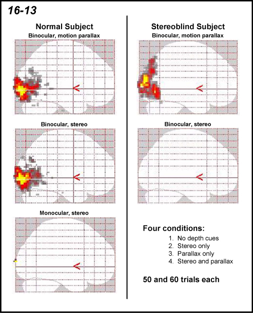
16-13 shows images obtained from a normal and a stereoblind subject demonstrating
the differences in brain activation. The images are a maximum intensity
projections showing a sagittal view of the two brains. For each display
shown in Figure 7, the scans collected for condition 1 (no depth) were
contrasted with the scans obtained with either condition 2 or 3 (disparity
cues only and motion parallax cues only). The top two figures show brain
activation for motion parallax under binocular viewing conditions. The
second two show the activation that occurs with disparity cues. There
was no activation in the brain of the stereoblind subject specific for
stereopsis. The bottom figure displays the control condition for the normal
subject showing no activation under monocular viewing conditions using
the displays with disparity. Figure 16-13 establishes that the procedures
we have devised work extremely well in specifically activating the brain
for stereopsis and motion parallax in the normal subjects and not activating
the brain in the stereoblind subjects. We have obtained similar results
in several other subjects. Figure
16-13 shows images obtained from a normal and a stereoblind subject demonstrating
the differences in brain activation. The images are a maximum intensity
projections showing a sagittal view of the two brains. For each display
shown in Figure 7, the scans collected for condition 1 (no depth) were
contrasted with the scans obtained with either condition 2 or 3 (disparity
cues only and motion parallax cues only). The top two figures show brain
activation for motion parallax under binocular viewing conditions. The
second two show the activation that occurs with disparity cues. There
was no activation in the brain of the stereoblind subject specific for
stereopsis. The bottom figure displays the control condition for the normal
subject showing no activation under monocular viewing conditions using
the displays with disparity. Figure 16-13 establishes that the procedures
we have devised work extremely well in specifically activating the brain
for stereopsis and motion parallax in the normal subjects and not activating
the brain in the stereoblind subjects. We have obtained similar results
in several other subjects.
|