![]()
So Lab - BEAM Bioinstrumentation Engineering Analysis and Microscopy
|
Research in the So Lab Lithographic Regulation of Cellular Migration and Rheology |
|||
|
|
Lithographic Control Of Cell Migration - We are developing a method to direct adherent cell migration based upon controlling cell polarization by asymmetric lithographic patterning of adhesion molecules. Microcontact printing of self-assembled monolayers (SAMs) of alkanethiolates on gold was used to generate a substrate consisting of serialized asymmetric teardrop patterns surrounded by non-adhesive regions. Cells plated on such substrates spread to take on the shape of the underlying pattern. Previous work by Whitesides and coworkers has demonstrated that cells on asymmetric patterns once released by electrochemical desorption move in the direction of polarization. We attempt to look at long range cell movement by creating a cellular ratchet - a pathway of teardrops through which the cell will migrate in a controlled fashion without a gradient of stimuli. Currently experiments are being conducted to determine the effect of teardrop aspect ratio and distance between patterns on the probability the cell will move towards the next pattern. We hypothesize that the forward-backward motion of these cells along this lithographic pathway follows a binomial statistics distribution and that the underlying physics of the situation is similar to that of a Brownian ratchet. We acknowledge support from NIHP01HL6485 and a NSF graduate research fellowship |
||
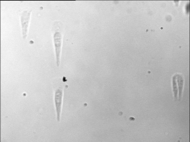 |
Figure 1: NIH3T3 fibroblasts plated on teardrop shaped patterns. Leica 10x objective. |
||
|
Lithographic Regulation Of Cellular Rheology - A single-pole magnetic trap in combination with two-photon fluorescence microscopy was used to determine the cytoskeletal stiffness and three-dimensional cytoskeletal structure of NIH 3T3 fibroblast cells plated on micropatterned substrates. Microcontact printing of self-assembled monolayers (SAMs) of alkanethiolates on gold was used to create substrates of islands surrounded by non-adhesive regions. The cells were physically constrained within nanometer high adhesive cylindrical posts of defined size on the surface of a titanium and gold coated coverslip. The islands were coated with the extracellular matrix protein fibronectin (FN) and a protein inhibiter was used to restrict cellular extension. After plating, the cells were fixed and stained with phalloidin. A high-speed, two-photon scanning microscope was used to resolve actin architecture in three dimensions and a fractal dimension measurement was performed to quantify the distribution of actin within the cell as a function of adhesion area. The experiments intend to test the hypothesis that cytoskeletal stiffness and structure is a function of adhesion area. It was discovered that larger patterns had a smaller fractal dimension. Microrheological experiments using a single pole magnetic trap are underway to quantify the storage and loss modulus of the cytoskeleton. We acknowledge support from NIHP01HL6485 and a NSF graduate research fellowship.
|
|||
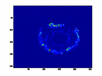 |
Figure 2: Two-photon image of a NIH3T3 fibroblast cell plated on a 40 um circular pattern. Cell was fixed with formalin and stained with AlexaFluor488-phalloidin. Image was taken at the widest cross-section of the cell. Zeiss 40x Fluar objective. |
||
[ So Lab - Mission Statement - Research - People - Documents - Facilities - Links - Resources - Sign-up Sheets ]
Comments about this webpage? Email us!