![]()
So Lab - BEAM Bioinstrumentation Engineering Analysis and Microscopy
|
Research in the So Lab Multifoci
Multiphoton Microscopy (MMM) |
||
|
|
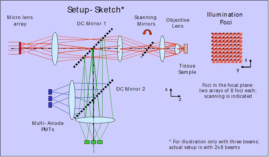
Investigators: Karsten Bahlmann, Timothy Ragan, Hyuk-Sang Kwon & Ki Hean Kim The combination of a multifocal multiphoton microscope (MMM) with a multi anode photon multiplier tube (PMT) enables time efficient three-dimensional (3D) fluorescence imaging of large tissue volumes at cellular resolution. The light from a 2 Watt Ti-sapphire laser, emitting ~150 fs pulses at 76 MHz, illuminates a micro lens array that passes 36 foci into the focal plane. The emission light is split in two colors, each equipped with one multi anode PMT adding to a total of 72 parallel detection channels. Each PMT in the multi anode PMT array corresponds to one illumination spot in the focal plane. As the foci are scanned the multi anode PMT readout is time correlated, generating images at up to 30 frames per second. The foci have a distance of 45 microns covering a total field of 270 microns X 270 microns.
Figure 1: Schematic of the optical setup of a multifoci multiphoton microscope (MMM).
|
|
 |
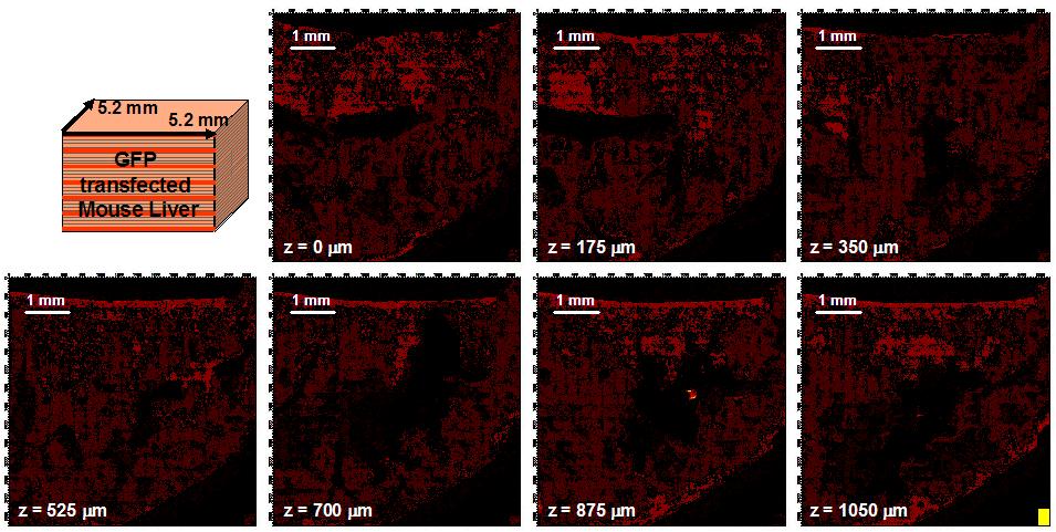
Figure
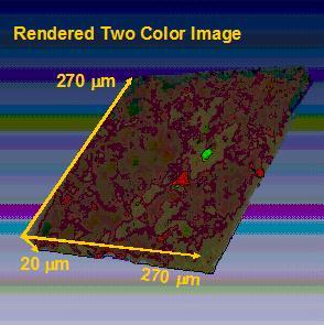
2: Two color image of Mouse Liver transfected with GFP cells - normalized. |
|
 |
||
|
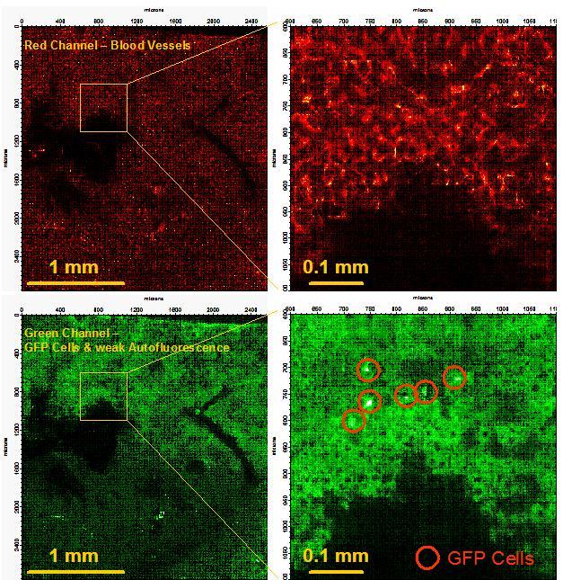
Figure 3: Study of the migration of cancer cells in tissue and whole organs by 3D-Cytometry - normalized. |
||
[ So Lab - Mission Statement - Research - People - Documents - Facilities - Links - Resources - Sign-up Sheets ]
Comments about this webpage? Email us!