![]()
So Lab - BEAM Bioinstrumentation Engineering Analysis and Microscopy
|
Research in the So Lab Tissue Spectroscopy |
||
|
|
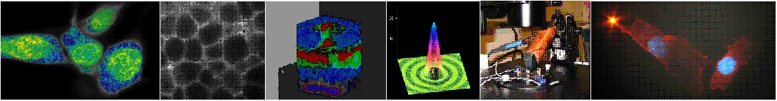
Tissue spectroscopy is a powerful method to identify and characterize endogenous fluorescence species. The relative abundance of these species is related to physiological and pathological states of the tissue. Fluorescence spectroscopy has been used to characterize different tissue types such as colon, lung, cervix, and skin. In addition to tissue type characterization, spectroscopy has been used to monitor the physiological state of a tissue. The differences in tissue excitation spectra has been used to for disease diagnosis such as distinguishing between malignant and normal tissues. |
|
|
|
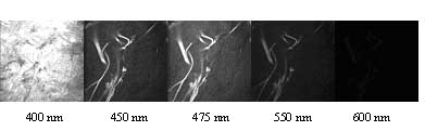
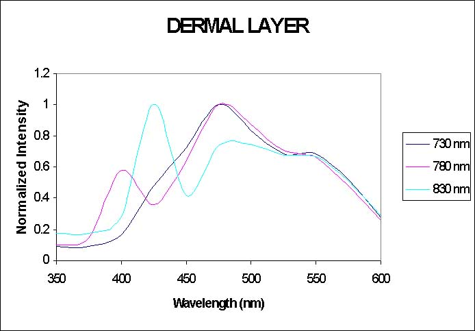
Figure 1. Representative spectral data and emission spectra for dermal layer in human skin. We have combined both two-photon imaging with spectroscopy in order to extract information about the chemical components contributing to the autofluorescence that we see in tissue. The analysis of tissue spectroscopic information with 3-D microscopic resolution has major advantages compared with typical tissue spectroscopy methods. Using different analysis techniques such as multivariate curve resolution, we are able to extract both the excitation and emission spectrum of the different components, such as collagen, elastin, NADH, melanin, and tryptophan. In addition, we are able to obtain the bulk emission spectrum and examine the spectrum from different layers and individual structures within the tissue. |
|
|
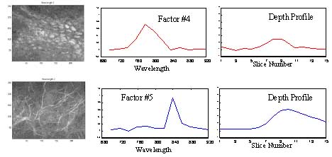
Figure 2. Representative spectral data, excitation spectra, and concentration profile for basal cells and elastin in human skin. |
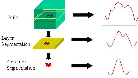
Figure 3. Schematic of bulk, layer, and structure resolved emission spectrum. |
|
[ So Lab - Mission Statement - Research - People - Documents - Facilities - Links - Resources - Sign-up Sheets ]
Comments about this webpage? Email us!