|


|
|||||||||||||||
Clinical light scattering spectroscopy imagining system for early cancer detection
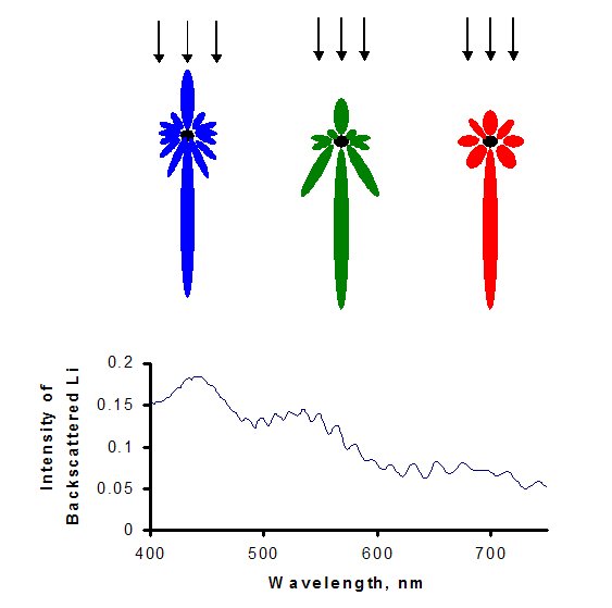
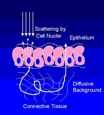
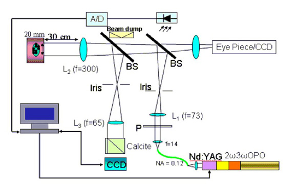
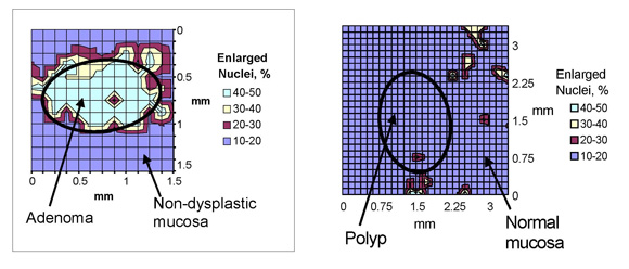
Background In the early stages of cancer development before becoming invasive, known as dysplasia and carcinoma in situ, the epithelial-cell morphology changes. In particular, the nuclei become enlarged, crowded, and hyperchromatic. The histological examinations are prone to inaccuracies in the location of biopsy. In this project, we utilize Imaging Light Scattering Spectroscopy (ILSS) to detect the morphological changes of epithelial cells such as nucleus enlargement. The diameter of non-dysplastic nuclei are in the range of 5-10um, where as dysplastic cells could be enlarged up to 20um across. The theory of ILSS is based on the prediction of the intensity of the backscattered light as a function of wavelength using Mie theory. This concept is demonstrated in Fig. 1. Based on the Mie theory, the backscattered pattern from a spherical particle can be theoretically calculated and it is a function of the size of the particle, the index of refraction contrast of the particle with its surrounding. In addition, as shown, the backscattered pattern is a function of wavelength and its largest component is in the direct backscattered direction. Therefore, in this imaging technique, we collect the direct backscattered wave and determine its variation as a function of wavelength, which once compared to the theory can be used to determine the particle size distribution. As the cancer is developed in the epithelial layer of the tissue, we would like to extract the size distribution of the nuclei in the top cell layer. However, the light penetrates through the top layer and is reflected from the lower tissues, which needs to be separated from the single direct backscattering. This is demonstrated in Fig 2. The multiple scattered beam as it reflects deeper from the tissue loses its polarization therefore having equal components in both parallel and perpendicular polarizations. Therefore, by subtracting the perpendicular polarization contribution from the parallel polarization, we can remove the multiple scattering signal. The experimental setup is shown in Fig 3. It consists of a light source, two 4f systems for collimation of the beam and collection of backscattered light and a polarizer and an analyzer to expose the image with a polarized beam and collect its contribution in two different polarizations. The light source is an optical parametric oscillator (OPO), the output of which is coupled to a fiber with the numerical aperture of 1.2. The OPO wavelength can be tuned between 410 to 680nm and it produces pulses of 5ns long at 20Hz with the energy per pulse ranging from 20 to 40 uJ. The light from the output of the fiber is collimated and then polarized through the polarizer P1. The polarized beam is then focused through a pinhole to select the collimation angle. The light from the pin hole is then collimated using lens L3, which combined with lens L2 determine the beam size diameter of the collimated beam (2 cm) which is used to expose the tissue. The backscattered light is focused again through a pinhole of about 5mm to collect angles of up to +/-0.5 degrees. The beam is imaged through lens L3, the output of which is passed through a birefringent material such as a calcite crystal where the two polarizations are separated and imaged on two different halves of the CCD. The perpendicular polarization is subtracted from the parallel polarization and the result is compared to Mie theory to determine the nucleus size distribution. The ex vivo colon tissue samples were obtained immediately after resection from patients undergoing coletomy. The Fig. 4 shows LSS images of two macroscopically similar colon polyps. The polyp on the left was identified as adenomatous by the histological diagnosis. The polyp on the right was classified as non-dysplastic inflammatory polyp. As outlined above, a series of polarized images at different wavelengths were taken, and the spectra were analyzed for each pixel of the imaged field. The parameters obtained were size and refractive index of the nuclei in each pixel. The field was divided in smaller 125 x 125 m2 regions and the percentage of nuclei larger than 10 micron was obtained for each of these areas. The resulting color-coded plots are shown. Figure on the left shows that, as expected, the nuclei are enlarged in the central, adenomatous region, but not in the surrounding tissue. For comparison, LSS image on the right shows that the spatial distribution of the nuclear sizes in the non-dysplastic polyp is uniform, with very few enlarged or hyperchromatic nuclei.
Current Work Recent Publications
|
|||||||||||||||

| [ Home | Overview | Research | People | Facilities | History | Events | Contact | MIT ] | Lab Intranet |