The Neural
Control of Visually Guided Eye Movements
C. Cortical Mechanisms of Visually Guided Saccadic Eye Movements
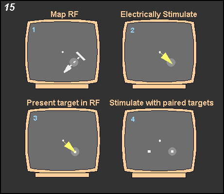 Figure
15 shows how the experiment was conducted: After advancing
a microelectrode into a given cortical area, the receptive field of the
neurons at the tip of the electrode was mapped out. This was accomplished
by moving a bar of light around while the monkey maintained fixation as
shown in #1 of this figure. We then electrically stimulated to generate
a saccadic eye movement as shown in #2. This shifted the center of gaze
into the receptive or motor field of the neurons stimulated. A target
was then placed at this site (#3); proper placement meant that the saccades
elicited by the target had the same vector as those elicited by electrical
stimulation. We then proceeded to present the paired targets with various
temporal asynchronies that were on some trials coupled with electrical
stimulation (#4). One of the targets was placed within the receptive or
motor field. Figure
15 shows how the experiment was conducted: After advancing
a microelectrode into a given cortical area, the receptive field of the
neurons at the tip of the electrode was mapped out. This was accomplished
by moving a bar of light around while the monkey maintained fixation as
shown in #1 of this figure. We then electrically stimulated to generate
a saccadic eye movement as shown in #2. This shifted the center of gaze
into the receptive or motor field of the neurons stimulated. A target
was then placed at this site (#3); proper placement meant that the saccades
elicited by the target had the same vector as those elicited by electrical
stimulation. We then proceeded to present the paired targets with various
temporal asynchronies that were on some trials coupled with electrical
stimulation (#4). One of the targets was placed within the receptive or
motor field.
|