
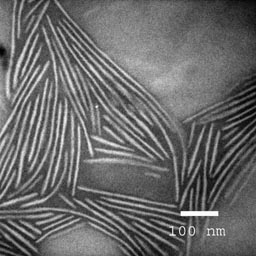
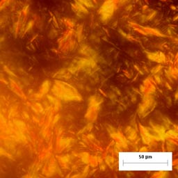

TEM of lamellar block copolymer nanostructures stained with osmium tetroxide (upper left). Polarized optical micrograph of a smectic liquid crystalline polymer (top). Cryo-TEM of a dilute polymer gel (right).
Microscopy allows direct visualization of polymer and protein nanostructures, including characterization individual defects, grain boundaries, or structures that may be challenging or impossible to detect by other means. Transmission electron microscopy (TEM) and cryo-TEM of vitrified samples allow nanoscale structures to be directly visualized in bulk materials, while atomic force microscopy (AFM) is used to characterize the height, nanostructure, and electrical properties in polymer thin films and interfaces (image on home page). Polarized optical microscopy techniques image patterns produced by birefringent liquid crystalline phases, allowing the liquid crystalline phase to be identified, and the interference between incident and reflected light creates color patterns in thin films that allow micron scale island or hole topographies to be imaged (image on home page).