So Lab - BEAM Bioinstrumentation
Engineering Analysis
and Microscopy
Research
in the So Lab
Core
Applications
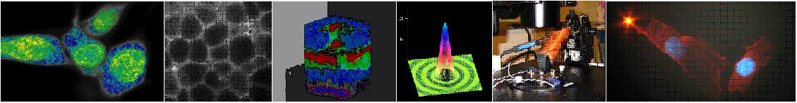
Carcinogenesis
Cancer
is caused by the accumulation of mutations in oncogenes and tumor
suppressor genes that control cell physiology and division. Mitotic
recombination has been estimated to be the underlying cause of LOH
25-50% of the time (Gupta et al., 1997; Morley et al., 1990; Zhu
et al., 1992). In collaboration with Prof. Bevin Engleward (BED,
MIT), we will combine genetic engineering with mechanico-optical
engineering to develop the technology to detect genetic instability
in mammals. A transgenic mouse will be engineered to carry a fluorescent
marker for identification of cells that have undergone a mitotic
recombination event. A high-throughput two-photon microscope system
will make it possible to quantify recombinant cells in situ in a
variety of cells, to characterize the cell types most prone to mitotic
recombination, and to discern the contribution of recombination
events that occur in stem cells. Yet another important application
will be in studying the effects of cancer chemotherapeutics on mitotic
recombination and in determining how specific genetic traits effect
cellular susceptibility to chemotherapy-induced mitotic recombination.
It is hoped that this line of research will ultimately aid in pharmacogenomics.
This new technology will be of fundamental importance in revealing
genetic and environmental processes that drive cancer-promoting
mitotic recombination events in mammals.
Optical
biopsy based on multi-photon imaging, spectroscopy and second harmonic
generation
Histological
analysis is the clinical standard for assessing tissue health and
the identification of pathological states. This analysis requires
tissues to be excised, fixated, sectioned, stained and subsequently
examined under a light microscope. The invasive nature of this process
justifies development of a supplementary approach without excision.
"Optical biopsy" is a promising alternative. Two-photon
microscope allows in vivo imaging of cellular and extracellular
matrix structures with sub-micron resolution inside intact 3-D tissue.
While the utility of two-photon microscopy for biological studies
has been clearly demonstrated in areas such as neurobiology and
embryology, its clinical potential remains unrealized. In collaboration
with Dr. Peter Kaplan (Unilever Edgewater Laboratory) and Dr. Christie
Ammirati (Pennsylvania State University Medical School) to address
this clinical need, we will develop and evaluate two-photon endoscopic
systems. We will characterize the performance this device in tissue
phantoms, animal models and excised human skin biopsy specimens.
Mechanotransduction
Mechanical
stimuli regulate many cellular responses, particularly within the
cardiovascular system. Understanding the process of mechanotransduction
has been challenging, in part because techniques for precisely controlled
mechanical stimulation have not been widely used. Under the leadership
of Prof. Roger Kamm (ME and DEB, MIT), we will develop rigorous,
highly specific and controllable methods of studying mechanotransduction
are needed based on novel micromanipulation techniques such as magnetic
trap, optical trap, and microlithography and cellular analysis tools
such as two-photon microscopy and spectroscopy, fluorescence based
laser tracking microrehology. In the first phase of this project,
we will investigate the mechanical responses of several novel genes
in smooth muscle cells and endothelial cells. This studies are highly
relevant to a number of cardiovascular diseases, including hypertensive
vasculopathy.
Torsional
effects on DNA-protein binding
In
collaboration with Prof. Peter Dedon (BED, MIT), we study the structural
effects and enzymological consequences of positive supercoiling
in DNA. Preliminary results reveal novel properties of helically
over-wound DNA consistent with an increase in the flipping of nucleobases
out of the helix. We propose to define the effects of positive supercoiling
on DNA structure and dynamics and on the activity of base-flipping
methyltransferase enzymes. Biochemical and spectroscopic characterization
in combination with biomechanical and biophysical methods will be
used to study the effect of positive supercoiling on DNA structure
and enzyme activity These complementary approaches will provide
important insights into the biological role of superhelical tension
in DNA physiology, DNA damage and DNA repair.