![]()
So Lab - BEAM Bioinstrumentation Engineering Analysis and Microscopy
|
Research in the So Lab Fiber 3-D Two-photon Endoscopy |
|
|
|
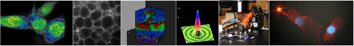
Two-photon fluorescence microscopic imaging has been demonstrated to be a new way to produce optical sectioned images in highly scattering medium such as tissues (ref.1 to 6). The development of a two-photon endoscope will be a critical step toward realizing non-invasive optical biopsy. It is clear that the imaging of intra-vessel structural morphology is of great clinical importance, especially the techniques that can image the tissue structural morphology beneath the surfaces of the internal biological vessels. However, the endoscope should be as small as possible because it would be used to image the internal human organ systems. A promising way is to use single-mode and multi-mode fiber as well as GRIN lens for light delivering, focusing, scanning and collecting. To do this, one should solved the following challenge problems.
(1) Group-Velocity Dispersion, |
.jpg) |
[ So Lab - Mission Statement - Research - People - Documents - Facilities - Links - Resources - Sign-up Sheets ]
Comments about this webpage? Email us!