![]()
So Lab - BEAM Bioinstrumentation Engineering Analysis and Microscopy
|
Research in the So Lab Two-photon Image Cytometry |
|
|
|
A
major direction we are pushing the application of two-photon microscopy
(TPM) is in the area of image cytometry. As the name implies, image cytometry
is an image-based study or measurement of cells. How image cytometry differs
from normal microscopic studies of cells is that very large populations
of cells (typically on the order of 104 to 108 cells) are imaged. To make
this technically feasible on the two-photon microscope, high-speed imaging
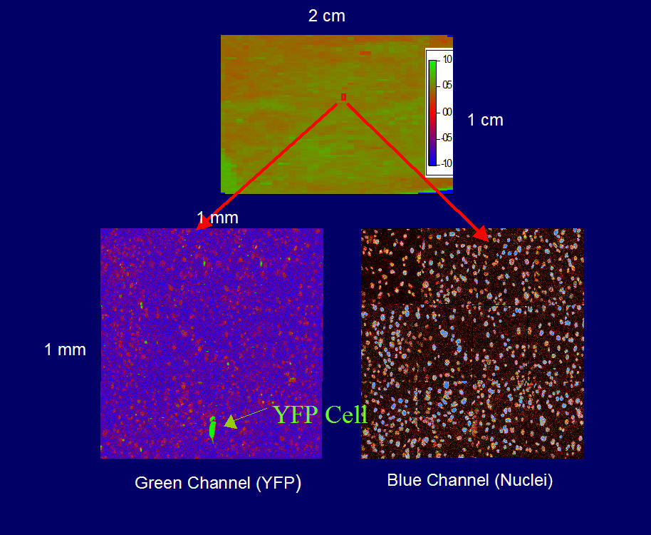
techniques are required. When we combine this with a mechanical stage that can translate the sample over several centimeters it becomes possible to image large areas in reasonable amounts of time. Below is a composite image of a population of a genetically modified 3T3 cells, which have had two non-functional yellow fluorescent protein (YFP) cassettes inserted into their genome, and a smaller zoomed-in region:
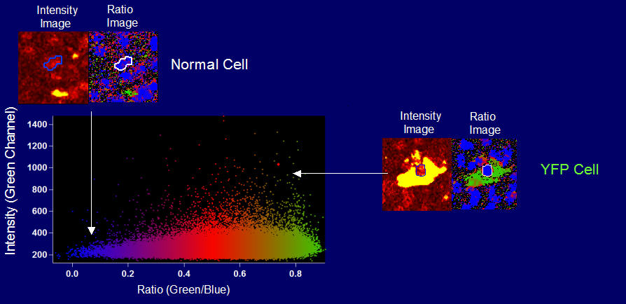
The cells have been stained with the DNA stain Hoescht 3328. The upper image is a ratio image of the green and blue channel, while the two lower images are the separated green channel and blue channels. One of the main strengths of image cytometry is its ability to identify rare events in a population of cells. The above image shows a cell that is fluorescent yellow that has undergone a recombination event that has restored the fluorescence of the YFP gene. Since this recombination event is a very low probability event (1 cell in 105) a large number of cells must be imaged in order to find such an event. We can also segment the cells and classify them using a cluster plot:
One
of the main strengths of TPM is its ability to image thick tissues specimens.
This gives us the ability to perform image cytometry in thick 3D samples
such as tissues. Below is a composite image of a ex vivo human skin sample
1 cm which has been imaged down to a depth of 70 microns. Thus it is possible
to perform image cytometry on cells while they are still in their intact
state, preserving many of their biochemical and mechanical inputs, and
most importantly their native 3D morphology and its relation to the 3D
architecture of the tissue. This provides a wealth of information about
tissue biophysics and biology on macroscopic samples that has not been
available before. We
are currently extending the capability of the instrument by increasing
the scanning speed to make it comparable to processing rates found in
flow cytometry, and combing histological sectioning to allow us to evaluate
specimens that have an axial extent greater than the standard 200 - 500
micron limit in two-photon microscopy. |
[ So Lab - Mission Statement - Research - People - Documents - Facilities - Links - Resources - Sign-up Sheets ]
Comments about this webpage? Email us!
.jpg)