![]()
So Lab - BEAM Bioinstrumentation Engineering Analysis and Microscopy
|
Research in the So Lab Fast Fluorescence Microrheology |
||
|
|
Investigator: Maxine Jonas To shed lights on mechanotransduction pathways in cardiovascular cells, we wish to explore the relationships between cellular rheology and both the recruitment of signaling molecules and the cytoskeletal remodeling by the cell. To that end, we have designed and built a novel microrheological device, a fluorescence laser tracking microrheometer (FLTM), capable of determining the stress/strain relationship of a sample at specific intracellular locations. This instrument expands upon the technology of laser tracking microrheometry, developed by Kuo et al. [1], to monitor the trajectory of a single fluorescent bead subjected to Brownian motion. Analysis based on the equipartition theorem subsequently allows for the extraction of the frequency-dependent complex shear modulus G*, index of the cell's resistance to deformation and fluid- vs. solid-like behavior. The FLTM (Figure 1) has a resolution of 5 nm over a frequency range extending from 1 Hz to 50 kHz. Additionally, our choice of fluorescent probes enables us to target cellular structures with molecular specificity. |
|
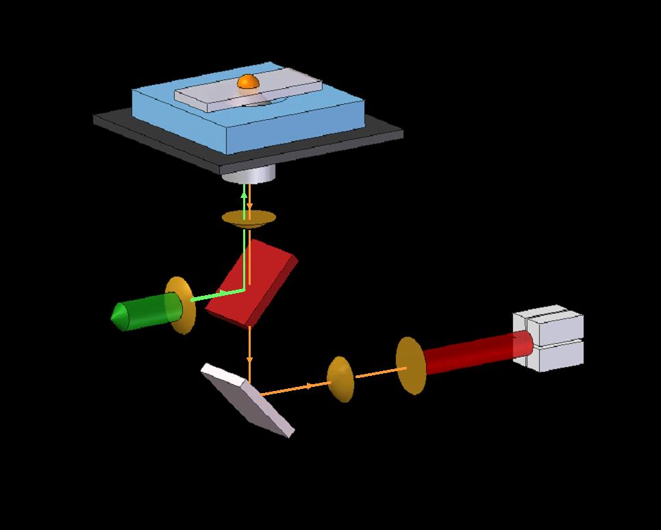 |
Figure 1 - Schematic representation of the fluorescence laser tracking microrheometer (FLTM). A laser beam is collimated through a custom light path and shone onto a 100-micron X 100-micron area of a sample positioned on the stage of an inverted microscope. The excited fluorescent bead contained in the illumination volume emits photons that are detected, after filtration and magnification, by a quadrant photomultiplier tube (PMT). Bead position signals are inferred at the PMT level from the difference in photocurrents generated by opposing pairs of quadrants, further amplified into digitized voltages by a four-channel analog-to-digital converter. |
|
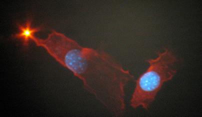 |
Figure 2 - Fibroblast cells (NIH 3T3) imaged after fixation and staining by Alexa Fluor-labeled phalloidin (F-actin) and by DAPI (nuclei). A 1-micron fluorescent polystyrene bead, coated with integrin antibodies, has attached to the cell membrane, and can be used for passive microrheology measurements. We have demonstrated the capabilities of the FLTM by obtaining good agreement with previously reported rheological measurements of polyacrylamide gels. In vivo, the FLTM is also a valuable tool (Figure 2) that enables the estimation of the fluid-like vs. solid-like properties of the cell cytoskeleton. |
|
|
Future
work includes 1. Yamada S., Wirtz D., Kuo S.C. (2000). Mechanics of living cells measured by laser tracking microrheology. Biophys. J., 78 (4): 1736-1747. |
||
[ So Lab - Mission Statement - Research - People - Documents - Facilities - Links - Resources - Sign-up Sheets ]
Comments about this webpage? Email us!