![]()
So Lab - BEAM Bioinstrumentation Engineering Analysis and Microscopy
|
Research in the So Lab Standing-wave Total Internal Reflection Fluorescence Microscopy (SW-TIRM) Microscopy for high resolution beyond the diffraction limit |
|||
|
|
Investigators: Euiheon Chung & Daekeun Kim Light microscopy is widely used in biomedical research to study living biological systems. A major limitation of optical imaging is its inability to resolve objects with separation below several hundred nanometers. Even though there are several scanning-probe methods with higher resolution such as atomic force microscopy, there is significant resolution degradation in soft biological specimens and this method is inherently slow due to point-by-point scanning. Thus there is a need to develop a wide-field optical imaging method with below 100nm resolution. |
||
|
To obtain lateral resolution beyond the diffraction limit in optical measurements, standing-wave total internal reflection fluorescence (SW-TIRF) microscopy has been developed. SW-TIRF uses the modulation of super-diffraction-limited evanescent excitation field to extract high spatial frequency content through the diffraction-limited optical imaging system. This system can obtain an effective point-spread function (PSF) with a full width at half maximum (FWHM) that is better than one sixth of the emission wavelength.
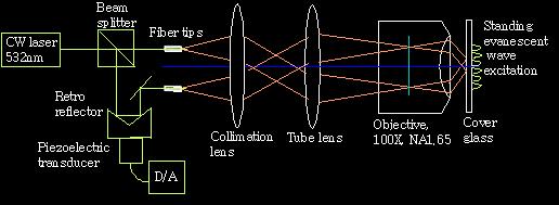
Fig.1 Schematic of objective-launch standing-wave total internal reflection fluorescence microscopy (SW-TIRF)
|
|
||
|
The use of standing evanescent wave imaging in a total internal reflection geometry have shown that lateral resolution better than 1/6 of emission wavelength can be achieved. The enhanced image results from the high-spatial frequency modulation to the conventional point-spread function. A
high NA objective lens-based standing wave total internal reflection fluorescence
microscopy has been developed. One dimensional lateral resolution improvement
using SW-TIRF is demonstrated with FWHM of about 100 nm that is approximately
one third of the diffraction limit [1].
|
|||
|
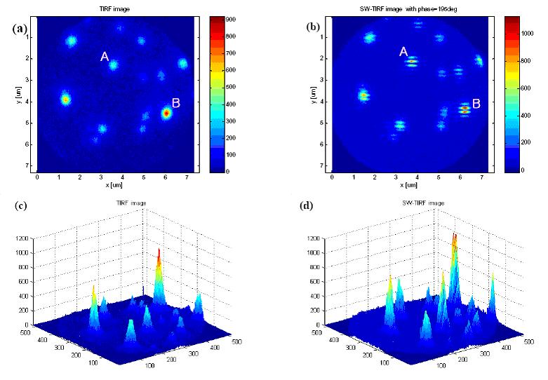
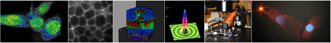
Fig. 2 Comparison of imaging 44 nm fluorescent beads (a) conventional TIRF and (b) SW-TIRF. (a), (b) Planar images using TIRF and SW-TIRF and (c), (d) corresponding intensity profiles. (The images were taken in the same setup while TIRF imaging was taken by blocking one excitation beam, A, B: regions of interest , NA=1.65, index of refraction of cover glass 1.788.) |
|
||
|
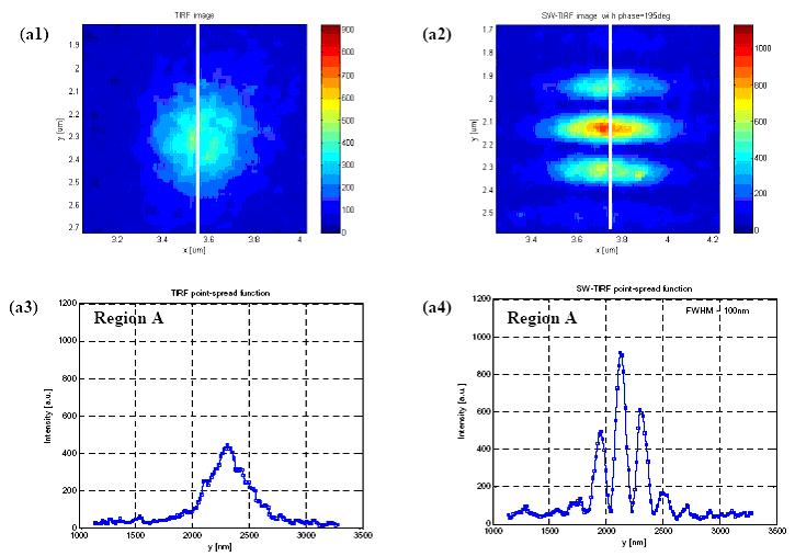
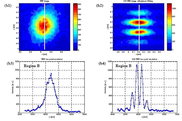
Fig. 3. (a1), (a3) and (b1), (b3) are the enlarged images and their intensity profiles in y-direction of region A under TIRF. (a2), (a4) and (b2), (b4), are the enlarged images and their intensity profiles in y-direction of region B under SW-TIRF.
1. E. Chung, D. Kim, and P.T.C. So, "Ultra-High Resolution Optical Imaging beyond the Diffraction Limit," The 2nd International Symposium on Nanomanufacturing, KAIST, Daejeon, Korea, Nov. 2004, pp. 158-163 |
|
||
[ So Lab - Mission Statement - Research - People - Documents - Facilities - Links - Resources - Sign-up Sheets ]
Comments about this webpage? Email us!