Tissue scanner for diagnosis of ex vivo surgical specimens


The objective of this instrument is to expand point based diffuse reflectance (DRS) and intrinsic fluorescence (IFS) to an imaging modality to provide fast, accurate diagnostic images of ex vivo tissue specimens during oncologic surgery, which together provide information on the metabolic, biochemical and morphological state of tissue that each modality alone cannot provide. By expanding our measurement from single points to imaging, we will be able to provide diagnostic images of tissue for more complete resection of diseased tissue and better conservation of normal tissue at cancer surgery. We will begin with an emphasis on diagnosis and imaging of breast cancer and anticipate expanding our studies to OTHER cancers.
We have constructed a bench-top DRS-IFS prototype scanner to scan large (up to 6x8 inches at 0.25mm resolution) tissue specimens. Two similar probes that are successfully employed in the probe-based systems will be used for the delivery and collection. One for diffusely reflected light, Xenon arc and other for IFS light source, pulsed diode pumped solid state laser that delivers 355 nm light pulses (SNV-40F-000, Teem Photonics) will be used and collected with miniature spectrometers, which later are digitally mapped to the same scanning area. The wide area imaging capability is achieved by mechanically scanning optical probes in an inverted geometry through a glass plate on which the specimen sits. The full mechanical scan of this area can be performed in less than 10 minutes by combining raster scanning of the stage and a series of triggers, but the final speed will depend upon the optical properties of breast tissue. Synchronization of translation control was done with a series of triggers for spectrograph acquisition during the scan. A computer is used to control data acquisition and analyze the spectroscopic data. The size and weight of the setup are 2x1x1 feet and 30 lbs, respectively, which could easily fit in an operating room.
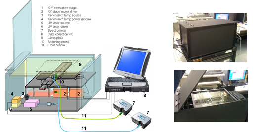
Figure 1.Shows the table top tissue scanner diagram (left) and the actual unit (top and bottom right).
In the past, using probe-based point measurements and DRS / IFS parameters, LBRC studies have successfully shown classification of normal breast tissue, fibrocystic change, fibroadenoma, and infiltrating ductal carcinoma. In a recent study of excised breast tissues from 17 patients, we acquired 202 spectra from 104 different sites using a portable clinical spectrometer and specially designed optical fiber probe, essentially identical to the optical fiber probe used in the spectroscopic scanner instrument proposed here. Spectroscopy results were compared to pathology diagnoses, and diagnostic algorithms developed based on parameters obtained via logistic regression with cross-validation. All invasive breast cancer specimens are correctly diagnosed. In addition to our results, multiple other publications have demonstrated the ability of DRS, IFS and similar optical modalities in diagnosis and classification of breast cancer in excised tissues. Collectively, these findings demonstrate and confirm our ability to detect and classify clinically significant breast lesions in excised tissue specimens, forming the basis for comprehensive margin assessment using tissue scanner under development.
|
 |


