|


|
 |
||||||
|
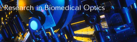
Research in Biomedical Optics and Spectroscopy Biomedical applications of lasers and laser spectroscopy are changing the face of medicine as it is currently practiced. The mission of the Laser Biomedical Research Center (LBRC), an NIH-sponsored research resource center, is to develop the scientific understanding required for advancing the applications of lasers in medicine and biology. Advanced spectroscopic methods are created for fundamental research involving biochemicals, cells, and ex vivo tissue samples to advance scientific understanding. Basic research serves as a foundation for instrumentation development and spectral model refinement that often results in the deployment of these techniques in clinical trials at medical centers and hospitals. Biomedical initiatives within the LBRC have resulted in the development of new spectroscopic methods to diagnose disease through minimally invasive procedures using absorption, fluorescence, Raman and intensity-based light scattering techniques; novel technologies for spectroscopic imaging of disease, in particular field-based light scattering techniques such as low-coherence interferometry and quantitative phase microscopy; and improved understanding and modeling of light transport in tissue. Basic studies in biophysics and biochemistry are also pursued to support the development of novel spectroscopic methods of disease diagnosis. Currently, the LBRC has technology research and development and collaborative projects in the following areas: A. Technology Research and Development (TDR) B. Collaborative Projects (CP) |
|||||||
| [ Home | Overview | Research | People | Facilities | History | Events | Contact | MIT ] | Lab Intranet |