|


|
|||||
|
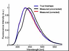
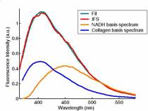
Intrinsic Fluorescence Spectroscopy (IFS) Intrinsic fluorescence spectroscopy (IFS) provides quantitative biochemical constituents. Key fluorescent tissue biochemicals include NADH, collagen crosslinks, tryptophan, FAD, and prophyrins. Fluorescence of these biochemicals is excited with laser light in the near UV to blue wavelengths and collected via a fiber optic probe (see fastEEM). A typical fluorescence emission spectrum is shown in Figure 1 (blue line). However, because tissue absorbs and scatters light, the measured emission spectrum can be highly distorted (Figure 1, black line). We employ a photon migration model[1, 2] to correct for these distortions and extract the true fluorescence emission spectrum (Figure 1, red line). This undistorted fluorescence spectrum is known as the intrinsic fluorescence spectrum.Subsequently, the intrinsic fluorescence spectrum is used to extract the basis biochemical components. For example, the two primary tissue fluorophores when excited with 340 nm light are collagen crosslinks and NADH. Their fluorescent spectra are shown in Figure 2 (blue and orange lines, respectively). A linear combination of these two basis spectra (Figure 2, green line) make up the intrinsic fluorescence spectrum measured from the tissue (Figure 2, red line). In this way, IFS quantitatively extracts the relative contributions of each biochemical.
|
|||||

| [ Home | Overview | Research | People | Facilities | History | Events | Contact | MIT ] | Lab Intranet |