Quantitative Microscopy and Tomography of Live Cells
Low-coherence diffraction Phase Microscopy
In phase shifting interferometry (PSI), the frequency of the reference beam is sinusoidally modulated and the phase information is extracted by acquiring and processing 3 or more interferograms. By contrast, Hilbert phase microscopy (HPM) offers much faster speed as it extracts sample phase information from a single interferogram by introducing spatial (phase-ramp) modulation between the reference and sample beams. In practice, both PSI and HPM use a separate reference beam for interference and extraction of the sample phase information. Therefore, for both techniques, the quality of the phase measurement may degrade due to mechanical vibrations in the setup, air flow in the beam paths, etc. Note that phase stability (or low phase noise) is critical for measurement of cell membrane fluctuations to assess membrane rheological properties or to study the correlation of phase fluctuations with electrical activity in mammalian cells (to be submitted). In 2006, core researchers at the LBRC developed a novel phase measurement technique known as diffraction phase microscopy (DPM) that minimizes system phase noise by using a common-path configuration.
Figure 1. Diffraction phase microscopy. VPS, virtual source point; G, grating; IP, image plane; L1,2 lenses (f1,2, respective focal distances); SF, spatial filter (expanded in the inset).
Figure 1.1 shows the schematic of the DPM setup, which illustrates the beam path after the sample plane. The transmitted light is separated into multiple copies of the original sample beam by using a diffraction grating placed in one of the intermediate planes. One of the diffracted beams passes through a pinhole that filters all the high spatial frequency information of the sample; the outcoming beam is a plane wave which is used as a reference beam. In such a configuration, the sample and the reference beams share most of the beam path; hence we call this a common path design, which ensures that the system phase noise is minimized. The data processing is similar to that in Hilbert phase microscopy.

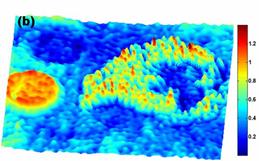 Figure 2. Quantitative phase image of human red blood cell. (b) Eosinophil chemotaxis approaching a red blood cell.
We have developed a low-coherence diffraction phase microscope with considerably lower phase noise that makes it useful for biological studies such as membrane electromotility in mammalian cells.
- T. Vo-Dinh, Biomedical Photonics Handbook (CRC Press, 2003).
- Park YK, Popescu G, Badizadegan K, Dasari RR, Feld MS. Diffraction phase and fluorescence microscopy. Optics Express 14, 8263-8 (2006) PMID: 19529201.
- Lue N, Choi W, Badizadegan K, Dasari RR, Feld MS and Popescu G. Confocal diffraction phase microscopy of live cells. Optics Letters 33, 2074-2076 (2008) PMCID: PMC2730468.
|
 |


