|


|
||||
|
Quantitative Microscopy and Tomography of Live Cells Tomographic Phase Microscopy The set up (Fig. 1) is based on a Mach-Zehnder heterodyne interferometer [5]. A helium-neon laser beam (l = 633 nm) is divided into sample and reference arm paths by a beam-splitter. A galvanometer-mounted tilting mirror is used to vary the angle of illumination of the sample, which is positioned between the oil-immersion condenser and objective lenses. In the reference arm, the laser beam passes through two acousto-optic modulators (AOMs), which shift the frequency of the laser beam by 1250 Hz. A beam-splitter recombines the sample and reference laser beams, forming an interference pattern at the image plane. With the speed 5000 frames per second, a CMOS camera (Photron 1024PCI) records 4 interferometric images for each illumination angle, from which one phase image can be calculated. To reconstruct a 3-D refractive index tomogram from the phase images, we first applied a procedure based on the filtered back-projection method [5], and then implemented the diffraction tomography [1]. The diffraction tomography handles diffraction from small organelles, and enables more accurate reconstruction throughout the entire 3-D volume. 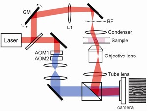 Figure 1. Tomographic phase microscope. GM: galvanometer scanning mirror; L1: f=250mm lens; BF: back focal plane of condenser lens; the frequency-shifted reference laser beam is shown in blue. To the right of the camera is a typical fringe pattern for a tilted beam illuminating a single HeLa cell [5]. Figures 2 and 3 show the reconstructed tomograms. A 3-D index tomogram for a single cell (Fig. 2 a,b) and x-y tomographic slices of the same cell at heights of z = 12, 9.5, 8.5, 7.5, 6.5 and 5.5 microns above the substrate (Fig. 2 c-h) show that the index of refraction is highly inhomogeneous, varying from 1.36 to 1.40. Bright field images for objective focus corresponding to Fig. 2 (e)-(f) are shown in Figure 2 (i)-(j), respectively. There is a clear correspondence between the tomographic and bright field images in terms of cell boundary, nuclear boundary, and size and shape of the nucleoli. 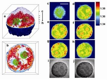 Figure 2. Refractive index tomogram of a HeLa cell. (a) 3-D rendered image. The outermost layer of the upper hemisphere of the cell is omitted to visualize the inner structure. Nucleoli are colored green and parts of cytoplasm with refractive index higher than 1.36 are colored red. The dotted box is a cube of side 20 mm. (b) Top view of (a). (c)-(h) Slices of the tomogram at heights indicated in (a). Scale bar, 10 mm. The color bar indicates the refractive index at l = 633 nm. (i) and (j) Bright field images for objective focus corresponding to (e) and (f), respectively. Note that the refractive index of the nucleus (n ≈1.36), apart from the nucleolus, is smaller than some parts of the cytoplasm (n≈1.36-1.39) and that the refractive index of the nucleoli, n≈1.38, is larger than that of the rest of the nucleus. This is contrary to the widely cited claims that the refractive index of the nucleus as a whole is higher than that of the rest of the cell. Similar results were obtained for cultured HEK 293 cells, B35 neuroblastoma cells, and primary rat hippocampal neurons. All cells imaged contained many small cytoplasmic particles with high refractive index, which may be lipid droplets, lysosomes, vacuoles, or other organelles. Assembling overlapping tomograms into a mosaic, tomographic phase microscopy can image a multi-cellular organism such as nematode C. elegans (Fig. 3). Several internal structures are clearly visible, including a prominent pharynx and digestive tract. 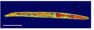 Figure 3. Mosaic of X-Y slices of index tomograms through the nematode C. elegans. Anterior is to the right. Scale bar, 50 mm. The color bar indicates the refractive index at λ = 633 nm. Illumination angles are limited to 60 degrees with respect to the optical axis by the numerical aperture of condenser and objective lenses. The missing angle produces an artifact, which can be suppressed by regularization techniques using various kinds of a priori knowledge. For example, we have shown that the positivity constraint can suppress the missing cone artifact by generating new data in the missing region [1]. We are developing an algorithm that can further suppress the missing cone artifact and increase the accuracy of refractive index prediction. Long-term observation of biological specimens is critical in many applications including cell cycle studies and monitoring the role of various chemicals inducing such effect as apoptosis in cancer cells. Label-free and 3-D imaging capability of TPM makes it highly appropriate for long-term observation of biological specimens because it is free from artifacts of the exogenous agents, which are invasive, and experience the problem of non-uniform binding and temporal degradation of signals. By installing a live-cell chamber, we are trying to develop an on-stage culture system that can be used for the tomographic phase microscopy. In addition, we are trying to speed up the data acquisition to minimize photo-induced damage on the biological specimen. This will extend the applicability of the instruments from observing the structure of biological specimens to studying their activities.
|
||||

| [ Home | Overview | Research | People | Facilities | History | Events | Contact | MIT ] | Lab Intranet |