|


|
||||
|
Quantitative Microscopy and Tomography of Live Cells Transmission-mode tomographic phase microscopy (TPM) has twice high transverse resolution than the diffraction limit, while its axial resolution is worse. This is mainly because of the limited angular coverage and the way we change the illumination direction with respect to a sample. Figure 1 shows the U-W cross-section of the transfer function for the scanning-beam type diffraction tomography in transmission and reflection mode. (U,W) is the spatial frequency components corresponding to (X,Z), in which X is one of the transverse coordinates and Z is the optical axis. Δax,tr is the axial resolution of the transmission mode diffraction tomography, and Δax,re is that of the reflection mode. The axial resolution of the reflection tomography is more than twice better than that of the transmission mode. 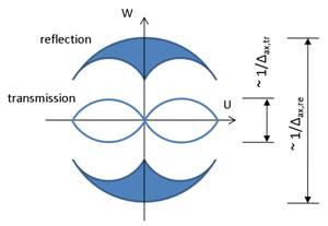 Figure 1. U-W cross section of the transfer function for transmission and reflection tomographic phase microscopy. The principle of reflection tomography has been known for a long time [1], but its experimental demonstration for a phase object has not been reported in the optical regime. It’s mainly because the reflections from multiple optical surfaces in the beam path easily overwhelm the signal from a sample. By effectively suppressing the reflection from the other surfaces than a sample, we are trying to implement the reflection tomography. By combining it with the transmission mode tomographic phase microscopy, we expect to provide better axial resolution and more accurate refractive index distribution of a sample.
|
||||

| [ Home | Overview | Research | People | Facilities | History | Events | Contact | MIT ] | Lab Intranet |