Imaging through Turbidity
Speckle-field based phase microscopy
We have developed several digital holographic microscopy techniques and utilizedthem to image the biological cells1-4. In an earlier work, we noted multiple scattering of light can be precisely measured as speckle electric field (E-field) and are deterministic3. Fig. 1B-C shows the amplitude and phase of the scattered field from a holographic diffuser using a Mach-Zenhder interferometry as shown in Fig. 1A. A thin sample, a polystyrene bead with 10 mm diameter was inserted after the diffuser and the E-fields are measured (Fig. 1D-E). Since the scattered fields are deterministic, we could retrieve the optical fields modified by the sample by dividing the scattered field with the sample by the scattered field without the sample (Figs. 1F-G). Removing singularity points by applying different speckle fields provides the sample induced field with high accuracy (Figs. 1H-I). The results indicate that we can quantitatively measure the deterministic scattered fields from turbid media using digital holographic technique.
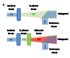
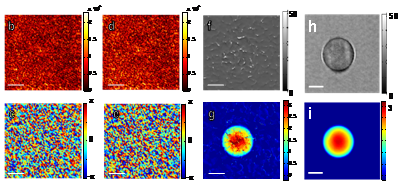
Figure 1.A-B.(a) Principle of speckle field measurement. H, holographic diffuser.S, sample.(b-c) amplitude (b) and phase (c) of the E-field without the sample,(d-e)amplitude (d) and phase (e) with the sample.(f-g)Amplitude (f) and phase (g) are images of the sample, a 10 mm polystyrene bead. (f-g)Amplitude (f) and phase (g) are images of the sample after removing singularity points. Scale bar, 5 μm. Colorbars indicate arbitrary units for the amplitude and radians for the phase, respectively. Images are adapted from Ref.3.
For the deterministic light scattering, there exists a relationship between an incident E-fields to the turbid medium and a scattered E-field at output, field transmission matrix. In order to study the light transport phenomena in complex media, we have measured the complete field transmission matrix of a scattering sample5. We have prepared a sheet of ZnOnano particles (d = 20±2 nm). From the E-field measurement at different incident beams, we have constructed the transfer matrix, which provides the full information on how light scatters in the sample. This information can be utilized to delivery optical information thought the complex medium or to image object embedded in the scattering tissues.
- Ikeda, T., Popescu, G., Dasari, R. R. & Feld, M. S. Hilbert phase microscopy for investigating fast dynamics in transparent systems. Opt. Lett.30, 1165-1168 (2005).
- Popescu, G., Ikeda, T., Dasari, R. R. & Feld, M. S. Diffraction phase microscopy for quantifying cell structure and dynamics. Opt. Lett.31, 775-777 (2006).
- Park, Y.-K. et al. Speckle-field digital holographic microscopy. Optics Express17, 12285-12292 (2009).
- Park, Y., Yamauchi, T., Choi, W., Dasari, R. & Feld, M. Spectroscopic phase microscopy for quantifying hemoglobin concentrations in intact red blood cells. Optics Letters34, 3668-3670 (2009).
- Park, Y.-K. et al. Direct measurement of eigen matrix of a scattering medium. (in preparation).
|
 |


