|


|
||||
|
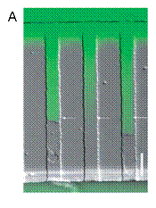
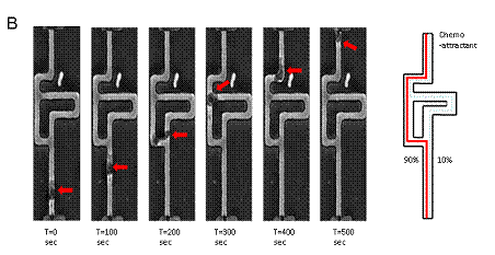
Collaborative Projects Spectroscopic monitoring of chemotaxis in microfluidic channels In collaboration with, Dr. Daniel Irimia, Assistant Professor, BioMEMS Resource Center at MGH and Harvard Medical School, we are developing several microfluidic devices capable of confining human neutrophils into small channels, for studying their migration in response to chemoattractants. The first generation of devices establishes the chemoattractant gradient along the channels by diffusion between a source and a sink, through the use of well balanced microfluidic streams of distinct fluids. The migration of cells through bifurcating channels are studied using similar devices, helping to quantify the bias in directional decision making in the presence of asymmetric stimulation of cells. Figure 1. Cell migration in microcapillaries. (A) A linear chemoattractant gradient is present in the empty channels and directs the migration of the HL60 cells (middle channel). After a cell enters one microchannel, a steep concentration difference is established between the front and back of the cell (top and bottom channels). (B) Arrows indicate that the fluorescence intensity is different depending on whether the channel is obstructed or not. Inside asymmetric mazes, human neutrophils chose the shorter path towards the source of fMLP 100 nM in the top channel in 90% of the observations (red outline).
|
||||

| [ Home | Overview | Research | People | Facilities | History | Events | Contact | MIT ] | Lab Intranet |