|


|
||||
|
Collaborative Projects Spectroscopic diagnosis and imaging of breast cancer Breast cancer is the leading cause of non-preventable cancer death among American women. More than 200,000 new cases are diagnosed each year, and more than 40,000 women die annually from breast malignancy. In collaboration with Maryann Fitzmaurice, MD at Case Western Reserve University, our efforts have been focused on developing spectroscopy instrumentations and techniques: diffuse reflectance (DRS), intrinsic fluorescence (IFS) and Raman for the diagnosis and imaging of breast cancer on two specific clinical applications: 1) guidance of stereotactic breast needle biopsy for mammographically detected microcalcifications: The study uses a clinical Raman system (compact Raman instrument and side-viewing Raman probes) specially designed and fabricated by the LBRC for use in conjunction with a commercial (ATEC) stereotactic breast needle biopsy system, in collaboration with Suros Surgical Systems, Inc., an interventional medical device manufacturer of minimally invasive surgical platform technologies for biopsy, tissue removal and biopsy site marking. This clinical Raman system is now located at University Hospitals-Case Medical Center (UH-CMC), where in vivo and ex vivo clinical studies are ongoing. 2) Intraoperative assessment of margins of resection and sentinel lymph nodes (LN) at breast cancer surgery, an important step in surgical management of breast cancer that has been inadequately addressed to date. This study is to apply DRS, IFS and Raman for intraoperative surgical margin and sentinel LN assessment to bench-top tissue scanners, as a clinical tool for ex vivo intraoperative assessment of the margins of breast specimens and sentinel LNs, to provide real-time feedback to the surgeon during breast cancer surgery. DRS, IFS and Raman spectroscopy are all being actively pursued as tools for the diagnosis of breast cancer, and advantageous for assessment of breast margins and sentinel LNs. Unlike traditional pathology diagnosis, spectroscopic diagnosis can be performed in real-time. Spectroscopic imaging techniques can examine the entire breast margin (Figure 1) and the entire capsular surface of the sentinel LN and, therefore, are not prone to sampling errors inherent in traditional pathology examination. Our spectroscopic techniques are also quantitative and therefore more objective than the traditional approach, which is subject to pathologist interpretation. This tissue scanner advances our probe-based technology from point measurements to a wide-field imaging regime more suitable for assessment of surgical margins and sentinel LNs. 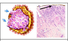 Figure 1. A: Diagram of positive breast surgical margin. B: Photomicrograph of breast cancer at an inked surgical margin (arrow; H&E; 20X). 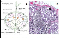 Figure 2. A: diagram of LN showing location of subcapsular sinus (A). B: photomicrograph of axillary LN with subcapsular breast cancer metastasis (arrow; H&E; 20X).
|
||||

| [ Home | Overview | Research | People | Facilities | History | Events | Contact | MIT ] | Lab Intranet |