Quantitative Phase Microscopy
Motivation
Quantitative phase imaging is a new optical approach that promises to revolutionize microscopy studies in cell biology. It provides biologists with a tool to study static and dynamic physical properties of live cells quantitatively, without exogenous contrast agents. For past several years, there has been considerable work on the development of quantitative phase microscopy (QPM) techniques at the G. R. Harrison Spectroscopy Laboratory; for example, the use of a harmonically-related pair of wavelengths and a phase-referenced interferometer. In the recent past, several QPM techniques have been developed that include: Fourier phase microscopy (FPM), Heterodyne Mach-Zehnder phase microscopy, Hilbert phase microscopy and Diffraction phase microscopy (DPM). These techniques have been employed to determine dry mass, refractive index of live cells and dynamic of cells such as cell’s membrane fluctuations and cell’s motion.
Fourier phase microscopy (FPM)
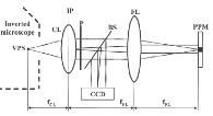
Fourier phase microscopy (FPM) combines a microscope with a phase shifting interferometer1. FPM is based on the principle of the interference of a scattered field and an unscattered field from a sample. The technique is inherently stable against phase noise because it does not require using two separate beams as in typical interferometry experiments. Phase shifting between scattered and unscattered fields, is accomplished by means of a programmable phase modulator (PPM, Hamamatsu Photonic, Japan) at the Fourier plane. The phase image of a sample is derived from a 4-frame phase shift algorithm. By replacing the PPM with a fast refresh rate liquid crystal modulator (Phase Contrast Filter (PCF), Hamamatsu, Japan), we can obtain images a rate of about 10Hz. Quantitative image of live cells and dynamic event of dissolving sugar crystal have been studied to demonstrate stability and acquisition speed.
|
Figure 1. Fourier phase microscopy setup |
Heterodyne Mach-Zehnder Phase Microscopy
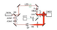
An alternative interferometric method to the common path geometry in FPM is to use frequency modulation, configured in Mach-Zehnder configuaration2. Two acousto-optic modulators (AOMs) were used in the reference beam path to introduce a series of phase shifts. The technique uses a high speed camera for recording the intensity images from which phase information can be retrieved. Our study of voltage-dependent cell motion has demonstrated the stability and speed of this approach.
|
Figure 2. Heterodyne Mach-Zehnder phase microscopy setup |
Hilbert Phase Microscopy (HPM)
HPM combines an inverted microscope built with a Mach-Zehnder interferometer3. Sample and reference arms are brought to interfere at the image plane of the microscope to form spatial interference patterns. These patterns are encoded with phase shift information of the sample. By extracting the sinusoidal term of the interferogram and applying the concept of analytic continuation to the spatially varying fields, the Hilbert transform relationship between the real and imaginary part of a complex analytic signals are used to obtain the phase shifts. In HPM, the data is acquired in a single shot, hence the phase imaging speed is limited only by the frame acquisition rate of the recording device (CCD) in contrast with FPM technique. HPM has more applicability to live cell studies as cell membrane dynamics2,4, cell-division cycle and organelle transport occur at shorter time scales. Several applications including refractive index and dry mass of cell, tissue refractive index and flow cytorefractometry are being studied with this HPM setup.
|
Figure 3. Hilbert phase microscopy setup |
Diffraction Phase Microscopy (DPM)
The best features of the FPM and HPM techniques have been combined to become another type of phase microscopy: Diffraction Phase Microscopy (DPM) 5,6. DPM uses the HPM interferometry technique to acquire a phase image in a single shot and uses the common path geometry of an FPM setup to obtain the highest stability. After the object beam passes through the sample in DPM it is diffracted by a phase grating into two diffraction orders. One of these is filtered with a spatial filter to generate a reference beam. The spatial filter consists of a pinhole placed at the Fourier plane to filter out high frequencies of spatial modulation. After traveling side by side through the same optical elements, the two beams are brought back to interfere at the image plane. HPM processing is then used to extract the phase image. DPM achieves the highest stability of all our techniques, and is currently being used to study the dynamics of red blood cells and neurons.
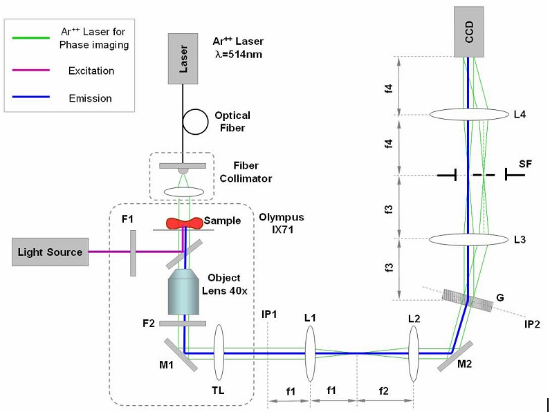
The DPM experimental setup is depicted in Fig. 4. An inverted microscope (IX71, Olympus Inc.) is equipped for standard epi-fluorescence, using a UV lamp and an excitation-emission filter pair, F1-F2. An Ar++ laser (λ = 514μm) is used as an illumination source for transmission phase imaging. Through its video output port, the microscope produces the image of the sample at the image plane IP1 with magnification M = 40. The lens system L1-L2 is used to collimate the unscattered field (spatial DC component) and further magnify the image by a factor f2/ f1=3, at the plane IP2. An amplitude grating G is placed at IP2, which generates multiple diffraction orders containing full spatial information about the sample image. The goal is to isolate the 0th and 1st orders and to create a common-path Mach-Zehnder interferometer, with the 0th order as the reference beam and the 1st order as the sample beam.
To accomplish this, a standard 4-f spatial filtering lens system L3-L4 is used. This system selects only the 0th and 1st order and generates the final interferogram at the CCD plane. The 0th order beam is low-pass filtered using a pinhole placed at the Fourier plane L3 so that it becomes a plane wave after passing through lens L4. The spatial filter allows passing the entire frequency information of the 1st order beam and blocks the high frequency information of the 0th beam.
|
Figure 4. DPM setup. F1, 2, filters; M1, 2, mirrors; L1-4 lenses (f1-4, respective focal lengths); G, grating; SF, spatial filter; IP1, 2, image planes; SF, spatial filter.
|
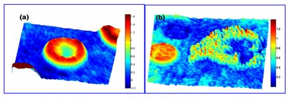
Due to its single shot nature, the DPM technique can be applied to investigating dynamic phenomena in live cells over temporal intervals that span from the millisecond scale or less to an entire cell cycle. We demonstrate this versatility with experiments of red blood cell (RBC) membrane fluctuations, which take place at the millisecond scale. We also illustrate imaging of white cell activity in blood smears, which develops over periods of minutes. Droplets of whole blood were placed between cover slips without further preparation. Figure 5(a) depicts an example of such quantitative phase image of a RBC. This shows the instant images of thermal fluctuations of the RBC membrane. This phenomenon of RBC “flickering” has been observed many years ago and intense efforts have been focused on understanding this dynamic process.
|
Figure 5. (a) Thermal fluctuations of red blood cell membrane. (b) Eosinophil chemotaxis and attack on red blood cell. Color bars represent optical phase in radians. |
Recent Publications
1. Popescu, G. et al. Fourier phase microscopy for investigation of biological structures and dynamics. Opt. Lett. 29, 2503-2505 (2004).
2. Fang-Yen, C. et al. Imaging voltage-dependent cell motions with heterodyne Mach-Zehnder phase microscopy. Opt. Lett. 32, 1572-1574 (2007).
3. Ikeda, T., Popescu, G., Dasari, R.R. & Feld, M.S. Hilbert phase microscopy for investigating fast dynamics in transparent systems. Opt. Lett. 30, 1165-1167 (2005).
4. Popescu, G. et al. Erythrocyte structure and dynamics quantified by Hilbert phase microscopy. J. Biomed. Opt. 10, 060503 (2005).
5. Park, Y.K., Popescu, G., Badizadegan, K., Dasari, R.R. & Feld, M.S. Diffraction phase and fluorescence microscopy. Opt. Exp. 14, 8263-8268 (2006).
6. Popescu, G., Ikeda, T., Dasari, R.R. & Feld, M.S. Diffraction phase microscopy for quantifying cell structure and dynamics. Opt. Lett. 31, 775-777 (2006).
7. Lue N, Choi W, Popescu G, Ikeda T, Badizadegan K, Dasari RR and Feld MS. Quantitative phase imaging of live cells using fast Fourier phase microscopy,
Appl. Opt., 46(10), 1836-42 (2007).
8. G. Popescu, Y. K. Park, N. Lue, L. Deflores, C. Best-Popescu, K. Badizadegan R. R. Dasari and M. S. Feld, “Optical imaging of cell mass and growth dynamics”, Am. J. Physiology, Cell Physiology 295, C538-542 (2008).
9. G. Popescu, Y. K. Park, W. Choi, R. R. Dasari, M. S. Feld and K. Badizadegan, “Imaging red blood cell dynamics by quantitative phase microscopy”, Blood Cells, Molecules and Diseases 41, 10-16 (2008).
|
 |