Tomographic phase microscopy and spring constants of cholesterol helical ribbons
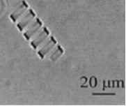
Self-assembly of helical ribbons in complex fluids is an interesting phenomenon, which poses fundamental questions about the molecular structure, elastic properties and kinetic evolution of these objects. In particular, solutions which contain cholesterol, non-ionic surfactants and lipids, spontaneously form helical ribbons with characteristic pitch angles of 11 and 54° (see Figure 1). It appears that the range of spring constants of our helices is such that they are suitable for measuring forces acting between nano-scale biological objects, such as antigen-antibody and enzyme-substrate interactions. In order to measure the forces, we need to know the spring constant of individual ribbons. We believe that the spring constants can be determined entirely by measuring the external dimensions of the helices. The dependence of the diameter of the ribbons on their thickness provides a crucial link between the elasticity and the external dimensions of the helices.
|
Figure 1. Phase contrast image of a helical ribbon |
The thickness of these cholesterol ribbons is less than 200 nm, which is below the diffraction-limit axial resolution of optical microscopes. However, the tomographic phase microscope recently developed at the MIT Spectroscopy Laboratory [2] is capable of determining the thickness with nanometer accuracy. The microscope uses interference between a sample beam and a reference beam to measure two-dimensional distributions of phase delays induced by a specimen in the sample beam.
The difference in phase across the field of view is due to the difference of refractive index between the ribbon and the media. The thickness t is deduced from the phase by means of the expression  , where , where  is the phase difference between the ribbon and the background, is the phase difference between the ribbon and the background,  nm is the wavelength of light and nm is the wavelength of light and  is the difference in refractive index between the ribbon and the medium. We have measured helical ribbons with diameters ranging from 15 to 140 micron. First results are consistent with expected diameter vs. thickness relation. is the difference in refractive index between the ribbon and the medium. We have measured helical ribbons with diameters ranging from 15 to 140 micron. First results are consistent with expected diameter vs. thickness relation.
These dramatic results demonstrate the ability of tomographic phase microscopy to measure biological structures well below the optical diffraction region, and illustrate its application to the problem of self assembly of helical cholesterol ribbons.
|
 |